Application
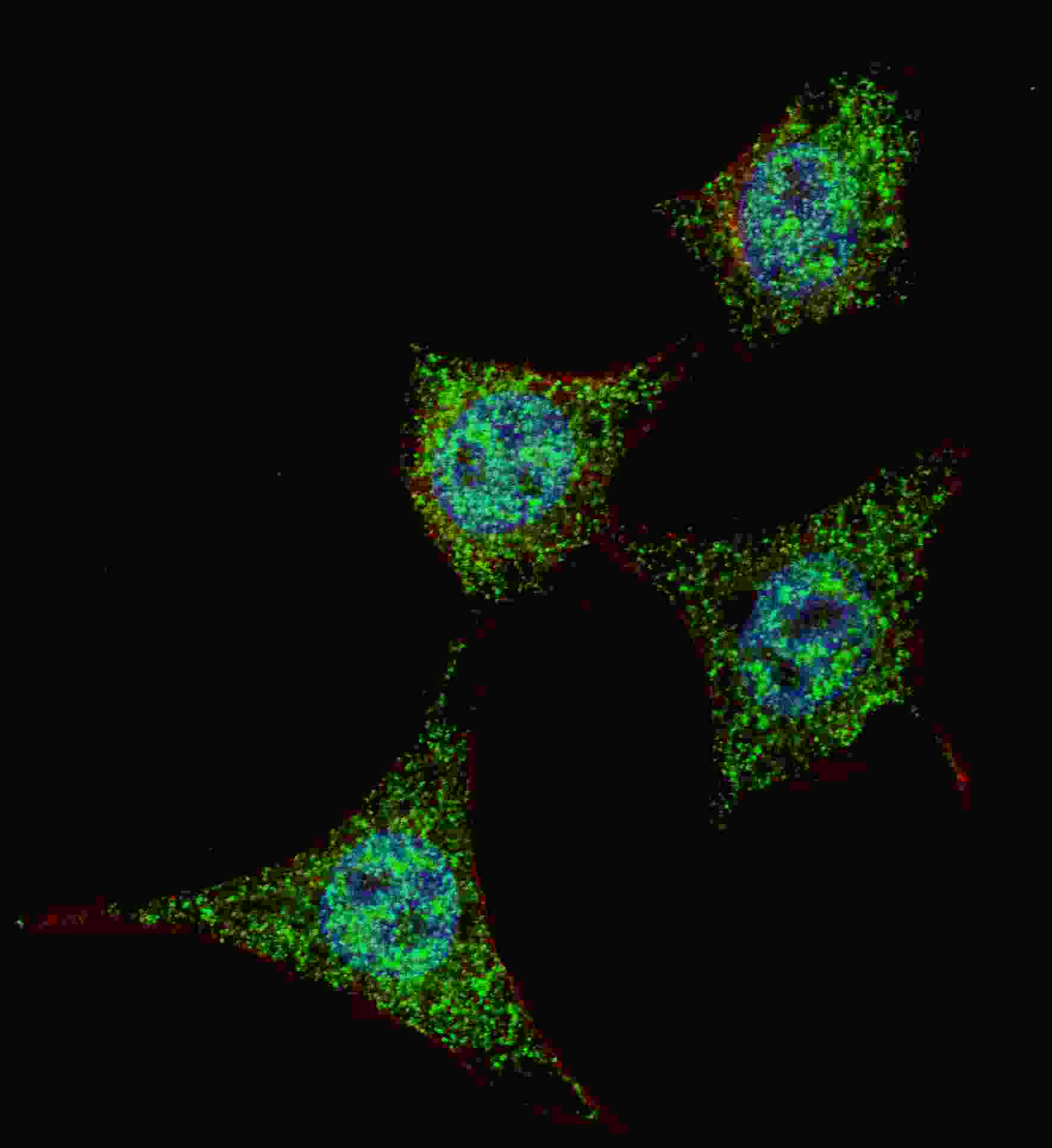
Fluorescent confocal image of HeLa cells stained with phospho-Bad-S99 antibody. HeLa cells were fixed with 4% PFA (20 min), permeabilized with Triton X-100 (0.2%, 30 min). Cells were then incubated with AP3039a phospho-Bad-S99 primary antibody (1:200, 2 h at room temperature). For secondary antibody, Alexa Fluor® 488 conjugated donkey anti-rabbit antibody (green) was used (1:1000, 1h). Nuclei were counterstained with Hoechst 33342 (blue) (10 μg/ml, 5 min). Note the highly specific localization of the phospho-Bad-S99 mainly to the cytoplasm.Application
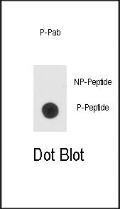
Dot blot analysis of anti-hBad-pS99 Phospho-specific Pab on nitrocellulose membrane. 50ng of Phospho-peptide or Non Phospho-peptide per dot were adsorbed. Antibody working concentrations are 0.5ug per ml.| Product Name | Phospho-Bad-S99 Antibody |
|---|---|
| Antibody Type | Primary Antibodies |
| Modification | p-S99 |
| Clonality | Polyclonal |
|---|---|
| Isotype | Ig |
| Host Species | Rabbit |
| Tested Applications | IFDB |
| IF:1:200 DB |
|
| Species Reactivity | Human |
| Concentration | 1mg/ml |
| Gene Synonyms | BBC6, BCL2L8 |
|---|---|
| Alternative Names | BAD BBC6 BCL2L8 Bcl2 antagonist of cell death Bcl-2-binding component 6 Bcl-2-like protein 8 Bcl-XL/Bcl-2-associated death promoter |
| Molecular Weight(MW) | 18392 Da |
| Function | Promotes cell death. Successfully competes for the binding to Bcl-X(L), Bcl-2 and Bcl-W, thereby affecting the level of heterodimerization of these proteins with BAX. Can reverse the death repressor activity of Bcl-X(L), but not that of Bcl-2 (By similarity). Appears to act as a link between growth factor receptor signaling and the apoptotic pathways |
| Tissue Specificity | Expressed in a wide variety of tissues. |
| Cellular Localization | Mitochondrion outer membrane. Cytoplasm. Note=Upon phosphorylation, locates to the cytoplasm |
| Entrez Gene | 572 |
|---|
Application
Fluorescent confocal image of HeLa cells stained with phospho-Bad-S99 antibody. HeLa cells were fixed with 4% PFA (20 min), permeabilized with Triton X-100 (0.2%, 30 min). Cells were then incubated with AP3039a phospho-Bad-S99 primary antibody (1:200, 2 h at room temperature). For secondary antibody, Alexa Fluor® 488 conjugated donkey anti-rabbit antibody (green) was used (1:1000, 1h). Nuclei were counterstained with Hoechst 33342 (blue) (10 μg/ml, 5 min). Note the highly specific localization of the phospho-Bad-S99 mainly to the cytoplasm.
Application
Dot blot analysis of anti-hBad-pS99 Phospho-specific Pab on nitrocellulose membrane. 50ng of Phospho-peptide or Non Phospho-peptide per dot were adsorbed. Antibody working concentrations are 0.5ug per ml.| Application Notes | IF:1:200 DB |
|---|
| Form | Liquid |
|---|---|
| Storage Instructions | For short-term storage, store at 4° C. For long-term storage, aliquot and store at -20ºC or below. Avoid multiple freeze-thaw cycles. |
| Storage Buffer | Purified polyclonal antibody supplied in PBS with 0.09% (W/V) sodium azide. This antibody is first purified by protein G affinity chromatography. Then, the antibody fraction is peptide affinity purified in a 2-step procedure with control and phosphorylated peptides. The phospho-specific antibody is eluted with high and low pH buffers and neutralized immediately, followed by dialysis against PBS. |


