Application
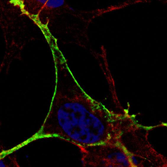
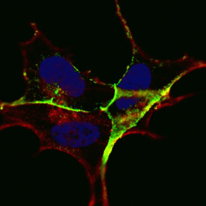
Fluorescent confocal image of SY5Y (SH-SY5Y; ATCC#CRL-2266) cells stained with AP3538a Phospho-KIT-Y721 antibody. SY5Y cells were fixed with 4% PFA (20 min), permeabilized with Triton X-100 (0.2%, 30 min), then incubated with AP3538a Phospho-KIT-Y721 primary antibody (1:100, 2 h at room temperature). For secondary antibody, Alexa Fluor® 488 conjugated donkey anti-rabbit antibody (green) was used (1:1000, 1h). Cytoplasmic actin was counterstained with Alexa Fluor® 555 (red) conjugated Phalloidin (5.25 μM, 25 min). Nuclei were counterstained with Hoechst 33342 (blue) (10 µg/ml, 3 min). Note the highly specific localization of the phosphorylated KIT immunosignal to the activated cell, and subcellularly to the membranes, especially the plasma membrane.Application
ibid. References for phosphorylated site Tyr-721: 1. Direct binding of CHK(SH2), FYN, PI3(PIK3R1) kinases [PubMed: 9038210]; 2. Tyrosine 721 is analogous to Y719 in the murine c-kit gene - a residue known to be essential for a normal spermatogonial proliferation. [PMID: 12322893]; 3. Most gastrointestinal stromal tumors (GISTs) show constitutive phosphorylation at Y721. [PMID: 15007386]; 4. Y721F mutant is less effective in driving cell proliferation, compared to wild type c-KIT. [PMID: 21887372]Application
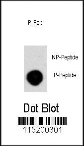
Dot blot analysis of anti-Phospho-KIT-Y721 Antibody on nitrocellulose membrane. 50ng of Phospho-peptide or Non Phospho-peptide per dot were adsorbed. Antibody working concentrations are 0.5ug per ml.| Product Name | Phospho-KIT-Y721 Antibody |
|---|---|
| Antibody Type | Primary Antibodies |
| Modification | p-Y721 |
| Clonality | Polyclonal |
|---|---|
| Isotype | Ig |
| Host Species | Rabbit |
| Tested Applications | IFDB |
| IF:1:10~50 DB |
|
| Species Reactivity | Human |
| Concentration | 1mg/ml |
| Gene Synonyms | SCFR |
|---|---|
| Alternative Names | KIT SCFR Mast/stem cell growth factor receptor Kit Piebald trait protein Proto-oncogene c-Kit Tyrosine-protein kinase Kit p145 c-kit v-kit Hardy-Zuckerman 4 feline sarcoma viral oncogene homolog CD_antigen=CD117 Flags: Precursor |
| Molecular Weight(MW) | 109865 Da |
| Function | Tyrosine-protein kinase that acts as cell-surface receptor for the cytokine KITLG/SCF and plays an essential role in the regulation of cell survival and proliferation, hematopoiesis, stem cell maintenance, gametogenesis, mast cell development, migration and function, and in melanogenesis. In response to KITLG/SCF binding, KIT can activate several signaling pathways Phosphorylates PIK3R1, PLCG1, SH2B2/APS and CBL. Activates the AKT1 signaling pathway by phosphorylation of PIK3R1, the regulatory subunit of phosphatidylinositol 3-kinase. Activated KIT also transmits signals via GRB2 and activation of RAS, RAF1 and the MAP kinases MAPK1/ERK2 and/or MAPK3/ERK1. Promotes activation of STAT family members STAT1, STAT3, STAT5A and STAT5B. Activation of PLCG1 leads to the production of the cellular signaling molecules diacylglycerol and inositol 1,4,5-trisphosphate. KIT signaling is modulated by protein phosphatases, and by rapid internalization and degradation of the receptor. Activated KIT promotes phosphorylation of the protein phosphatases PTPN6/SHP-1 and PTPRU, and of the transcription factors STAT1, STAT3, STAT5A and STAT5B. Promotes phosphorylation of PIK3R1, CBL, CRK (isoform Crk-II), LYN, MAPK1/ERK2 and/or MAPK3/ERK1, PLCG1, SRC and SHC1 |
| Tissue Specificity | Isoform 1 and isoform 2 are detected in spermatogonia and Leydig cells. Isoform 3 is detected in round spermatids, elongating spermatids and spermatozoa (at protein level). Widely expressed. Detected in the hematopoietic system, the gastrointestinal system, in melanocytes and in germ cells |
| Cellular Localization | Isoform 1: Cell membrane; Single-pass type I membrane protein Isoform 3: Cytoplasm. Note=Detected in the cytoplasm of spermatozoa, especially in the equatorial and subacrosomal region of the sperm head |
| Entrez Gene | 3815 |
|---|
Application
Fluorescent confocal image of SY5Y (SH-SY5Y; ATCC#CRL-2266) cells stained with AP3538a Phospho-KIT-Y721 antibody. SY5Y cells were fixed with 4% PFA (20 min), permeabilized with Triton X-100 (0.2%, 30 min), then incubated with AP3538a Phospho-KIT-Y721 primary antibody (1:100, 2 h at room temperature). For secondary antibody, Alexa Fluor® 488 conjugated donkey anti-rabbit antibody (green) was used (1:1000, 1h). Cytoplasmic actin was counterstained with Alexa Fluor® 555 (red) conjugated Phalloidin (5.25 μM, 25 min). Nuclei were counterstained with Hoechst 33342 (blue) (10 µg/ml, 3 min). Note the highly specific localization of the phosphorylated KIT immunosignal to the activated cell, and subcellularly to the membranes, especially the plasma membrane.
Application
ibid. References for phosphorylated site Tyr-721: 1. Direct binding of CHK(SH2), FYN, PI3(PIK3R1) kinases [PubMed: 9038210]; 2. Tyrosine 721 is analogous to Y719 in the murine c-kit gene - a residue known to be essential for a normal spermatogonial proliferation. [PMID: 12322893]; 3. Most gastrointestinal stromal tumors (GISTs) show constitutive phosphorylation at Y721. [PMID: 15007386]; 4. Y721F mutant is less effective in driving cell proliferation, compared to wild type c-KIT. [PMID: 21887372]
Application
Dot blot analysis of anti-Phospho-KIT-Y721 Antibody on nitrocellulose membrane. 50ng of Phospho-peptide or Non Phospho-peptide per dot were adsorbed. Antibody working concentrations are 0.5ug per ml.| Application Notes | IF:1:10~50 DB |
|---|
| Form | Liquid |
|---|---|
| Storage Instructions | For short-term storage, store at 4° C. For long-term storage, aliquot and store at -20ºC or below. Avoid multiple freeze-thaw cycles. |
| Storage Buffer | Purified polyclonal antibody supplied in PBS with 0.09% (W/V) sodium azide. This antibody is purified through a protein A column, followed by two-step phosphospecific peptide affinity purification. |


