Application
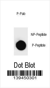
Dot blot analysis of Phospho-PARP1-T368 Antibody Phospho-specific Pab on nitrocellulose membrane. 50ng of Phospho-peptide or Non Phospho-peptide per dot were adsorbed. Antibody working concentrations are 0.6ug per ml.| Product Name | Phospho-PARP1-T368 |
|---|---|
| Antibody Type | Primary Antibodies |
| Antigen Alias | PARP1; ADPRT; PPOL; Poly [ADP-ribose] polymerase 1; ADP-ribosyltransferase diphtheria toxin-like 1; NAD(+) ADP-ribosyltransferase 1; Poly[ADP-ribose] synthase 1 |
| Modification | Phosphoralated |
| Clonality | Polyclonal |
|---|---|
| Isotype | Ig |
| Host Species | Rabbit |
| Tested Applications | DB |
| DB:1:500 |
|
| Species Reactivity | Human |
| Concentration | 1mg/ml |
| Gene Synonyms | ADPRT PPOL |
|---|---|
| Alternative Names | PARP1 ADPRT PPOL Poly [ADP-ribose] polymerase 1 ADP-ribosyltransferase diphtheria toxin-like 1 NAD(+) ADP-ribosyltransferase 1 Poly[ADP-ribose] synthase 1 |
| Molecular Weight(MW) | 113084 Da |
| Function | Involved in the base excision repair (BER) pathway, by catalyzing the poly(ADP-ribosyl)ation of a limited number of acceptor proteins involved in chromatin architecture and in DNA metabolism. This modification follows DNA damages and appears as an obligatory step in a detection/signaling pathway leading to the reparation of DNA strand breaks. Mediates the poly(ADP- ribosyl)ation of APLF and CHFR. Positively regulates the transcription of MTUS1 and negatively regulates the transcription of MTUS2/TIP150. With EEF1A1 and TXK, forms a complex that acts as a T-helper 1 (Th1) cell-specific transcription factor and binds the promoter of IFN-gamma to directly regulate its transcription, and is thus involved importantly in Th1 cytokine production |
| Tissue Specificity | This Phospho-PARP1-T368 Antibody is generated from rabbits immunized with a KLH conjugated synthetic phosphopeptide corresponding to amino acid residues surrounding T368 of human PARP1. |
| Cellular Localization | Nucleus. Nucleus, nucleolus. |
| Entrez Gene | 142 |
|---|---|
| Nucleotide Accession | NP_001609.2 |
Application
Dot blot analysis of Phospho-PARP1-T368 Antibody Phospho-specific Pab on nitrocellulose membrane. 50ng of Phospho-peptide or Non Phospho-peptide per dot were adsorbed. Antibody working concentrations are 0.6ug per ml.| Application Notes | DB:1:500 |
|---|
| Form | Liquid |
|---|---|
| Storage Instructions | For short-term storage, store at 4° C. For long-term storage, aliquot and store at -20ºC or below. Avoid multiple freeze-thaw cycles. |
| Storage Buffer | Purified polyclonal antibody supplied in PBS with 0.09% (W/V) sodium azide. This antibody is purified through a protein A column, followed by two-step phosphospecific peptide affinity purification. |


 Data sheet for OM217605