Application
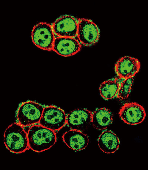
Fluorescent confocal image of HeLa cells stained with phospho-SMAD3-S208 antibody. HeLa cells were fixed with 4% PFA (20 min), permeabilized with Triton X-100 (0.2%, 30 min). Cells were then incubated with AP3249a phospho-SMAD3-S208 primary antibody (1:200, 2 h at room temperature). For secondary antibody, Alexa Fluor® 488 conjugated donkey anti-rabbit antibody (green) was used (1:1000, 1h). Cytoplasmic actin was counterstained with Alexa Fluor® 555 (red) conjugated Phalloidin (5.25 μM, 25 min). Pictures were taken on a Biorevo microscope (BZ-900, Keyence).Note the highly specific localization of the phospho-SMAD3 mainly to the nucleus, supported by Human Protein Atlas Data (http://www.proteinatlas.org/ENSG00000166949).Application
Confocal immunofluorescent analysis of Phospho-SMAD3-S208 Antibody(Cat#AP3249a) with Hela cell followed by Alexa Fluor 488-conjugated goat anti-rabbit lgG (green). Actin filaments have been labeled with Alexa Fluor 555 phalloidin (red).Application
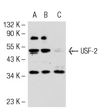
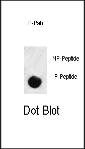
Dot blot analysis of anti-hSMAD3-S208 Phospho-specific Pab on nitrocellulose membrane. 50ng of Phospho-peptide or Non Phospho-peptide per dot were adsorbed. Antibody working concentrations are 0.5ug per ml.| Product Name | Phospho-SMAD3-S208 Antibody |
|---|---|
| Antibody Type | Primary Antibodies |
| Antigen Alias | SMAD3; MADH3; Mothers against decapentaplegic homolog 3; JV15-2; SMAD family member 3 |
| Modification | p-S208 |
| Clonality | Polyclonal |
|---|---|
| Isotype | Ig |
| Host Species | Rabbit |
| Tested Applications | IFDB |
| IF:1:10~50 DB |
|
| Species Reactivity | Human |
| Concentration | 1mg/ml |
| Gene Synonyms | MADH3 |
|---|---|
| Alternative Names | SMAD3 MADH3 Mothers against decapentaplegic homolog 3 JV15-2 SMAD family member 3 |
| Molecular Weight(MW) | 48081 Da |
| Function | Receptor-regulated SMAD (R-SMAD) that is an intracellular signal transducer and transcriptional modulator activated by TGF-beta (transforming growth factor) and activin type 1 receptor kinases. Binds the TRE element in the promoter region of many genes that are regulated by TGF-beta and, on formation of the SMAD3/SMAD4 complex, activates transcription Also can form a SMAD3/SMAD4/JUN/FOS complex at the AP-1/SMAD site to regulate TGF-beta-mediated transcription. Has an inhibitory effect on wound healing probably by modulating both growth and migration of primary keratinocytes and by altering the TGF- mediated chemotaxis of monocytes. This effect on wound healing appears to be hormone-sensitive. Regulator of chondrogenesis and osteogenesis and inhibits early healing of bone fractures (By similarity). Positively regulates PDPK1 kinase activity by stimulating its dissociation from the 14-3-3 protein YWHAQ which acts as a negative regulator |
| Tissue Specificity | This Phospho-SMAD3-S208 antibody is generated from rabbits immunized with a KLH conjugated synthetic phosphopeptide corresponding to amino acid residues surrounding S208 of human SMAD3. |
| Cellular Localization | Cytoplasm. Nucleus. Note=Cytoplasmic and nuclear in the absence of TGF-beta. On TGF-beta stimulation, migrates to the nucleus when complexed with SMAD4. Through the action of the phosphatase PPM1A, released from the SMAD2/SMAD4 complex, and exported out of the nucleus by interaction with RANBP1. Co-localizes with LEMD3 at the nucleus inner membrane MAPK-mediated phosphorylation appears to have no effect on nuclear import. PDPK1 prevents its nuclear translocation in response to TGF-beta |
| Entrez Gene | 4088 |
|---|
Application
Fluorescent confocal image of HeLa cells stained with phospho-SMAD3-S208 antibody. HeLa cells were fixed with 4% PFA (20 min), permeabilized with Triton X-100 (0.2%, 30 min). Cells were then incubated with AP3249a phospho-SMAD3-S208 primary antibody (1:200, 2 h at room temperature). For secondary antibody, Alexa Fluor® 488 conjugated donkey anti-rabbit antibody (green) was used (1:1000, 1h). Cytoplasmic actin was counterstained with Alexa Fluor® 555 (red) conjugated Phalloidin (5.25 μM, 25 min). Pictures were taken on a Biorevo microscope (BZ-900, Keyence).Note the highly specific localization of the phospho-SMAD3 mainly to the nucleus, supported by Human Protein Atlas Data (http://www.proteinatlas.org/ENSG00000166949).
Application
Confocal immunofluorescent analysis of Phospho-SMAD3-S208 Antibody(Cat#AP3249a) with Hela cell followed by Alexa Fluor 488-conjugated goat anti-rabbit lgG (green). Actin filaments have been labeled with Alexa Fluor 555 phalloidin (red).
Application
Dot blot analysis of anti-hSMAD3-S208 Phospho-specific Pab on nitrocellulose membrane. 50ng of Phospho-peptide or Non Phospho-peptide per dot were adsorbed. Antibody working concentrations are 0.5ug per ml.| Application Notes | IF:1:10~50 DB |
|---|
| Form | Liquid |
|---|---|
| Storage Instructions | For short-term storage, store at 4° C. For long-term storage, aliquot and store at -20ºC or below. Avoid multiple freeze-thaw cycles. |
| Storage Buffer | Purified polyclonal antibody supplied in PBS with 0.09% (W/V) sodium azide. This antibody is first purified by protein G affinity chromatography. Then, the antibody fraction is peptide affinity purified in a 2-step procedure with control and phosphorylated peptides. The phospho-specific antibody is eluted with high and low pH buffers and neutralized immediately, followed by dialysis against PBS. |


 Data sheet for OM217736