Application
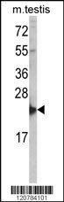
Western blot analysis of RAN Antibody (Center) in mouse testis tissue lysates (35ug/lane). RAN (arrow) was detected using the purified Pab.Application
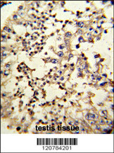
Formalin-fixed and paraffin-embedded human testis reacted with RAN Antibody (Center), which was peroxidase-conjugated to the secondary antibody, followed by DAB staining. This data demonstrates the use of this antibody for immunohistochemistry; clinical relevance has not been evaluated.Application
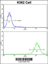
Flow cytometric analysis of K562 cells using RAN Antibody (Center)(bottom histogram) compared to a negative control cell (top histogram)FITC-conjugated goat-anti-rabbit secondary antibodies were used for the analysis.Application
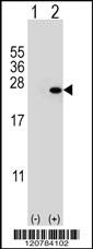
Western blot analysis of RAN (arrow) using rabbit polyclonal RAN Antibody (Center) . 293 cell lysates (2 ug/lane) either nontransfected (Lane 1) or transiently transfected (Lane 2) with the RAN gene.| Product Name | RAN Antibody (Center) |
|---|---|
| Antibody Type | Primary Antibodies |
| Antigen Alias | RAN; ARA24; GTP-binding nuclear protein Ran; Androgen receptor-associated protein 24; GTPase Ran; Ras-like protein TC4; Ras-related nuclear protein |
| Clonality | Polyclonal |
|---|---|
| Isotype | Ig |
| Host Species | Rabbit |
| Tested Applications | WBIHCFC |
| WB:1:50~100 IHC |
|
| Species Reactivity | Human |
| Concentration | 1mg/ml |
| Gene Synonyms | ARA24 |
|---|---|
| Alternative Names | RAN ARA24 GTP-binding nuclear protein Ran Androgen receptor-associated protein 24 GTPase Ran Ras-like protein TC4 Ras-related nuclear protein |
| Molecular Weight(MW) | 24423 Da |
| Function | GTP-binding protein involved in nucleocytoplasmic transport. Required for the import of protein into the nucleus and also for RNA export. Involved in chromatin condensation and control of cell cycle (By similarity). The complex with BIRC5/ survivin plays a role in mitotic spindle formation by serving as a physical scaffold to help deliver the RAN effector molecule TPX2 to microtubules. Acts as a negative regulator of the kinase activity of VRK1 and VRK2 |
| Tissue Specificity | Expressed in a variety of tissues. |
| Cellular Localization | Nucleus. Cytoplasm. Melanosome. Note=Predominantly nuclear during interphase (By similarity) Becomes dispersed throughout the cytoplasm during mitosis Identified by mass spectrometry in melanosome fractions from stage I to stage IV |
| Entrez Gene | 5901 |
|---|
Application
Western blot analysis of RAN Antibody (Center) in mouse testis tissue lysates (35ug/lane). RAN (arrow) was detected using the purified Pab.
Application
Formalin-fixed and paraffin-embedded human testis reacted with RAN Antibody (Center), which was peroxidase-conjugated to the secondary antibody, followed by DAB staining. This data demonstrates the use of this antibody for immunohistochemistry; clinical relevance has not been evaluated.
Application
Flow cytometric analysis of K562 cells using RAN Antibody (Center)(bottom histogram) compared to a negative control cell (top histogram)FITC-conjugated goat-anti-rabbit secondary antibodies were used for the analysis.
Application
Western blot analysis of RAN (arrow) using rabbit polyclonal RAN Antibody (Center) . 293 cell lysates (2 ug/lane) either nontransfected (Lane 1) or transiently transfected (Lane 2) with the RAN gene.| Application Notes | WB:1:50~100 IHC |
|---|
| Form | Liquid |
|---|---|
| Storage Instructions | For short-term storage, store at 4° C. For long-term storage, aliquot and store at -20ºC or below. Avoid multiple freeze-thaw cycles. |
| Storage Buffer | Purified polyclonal antibody supplied in PBS with 0.09% (W/V) sodium azide. This antibody is prepared by Saturated Ammonium Sulfate (SAS) precipitation followed by dialysis against PBS. |


