Application
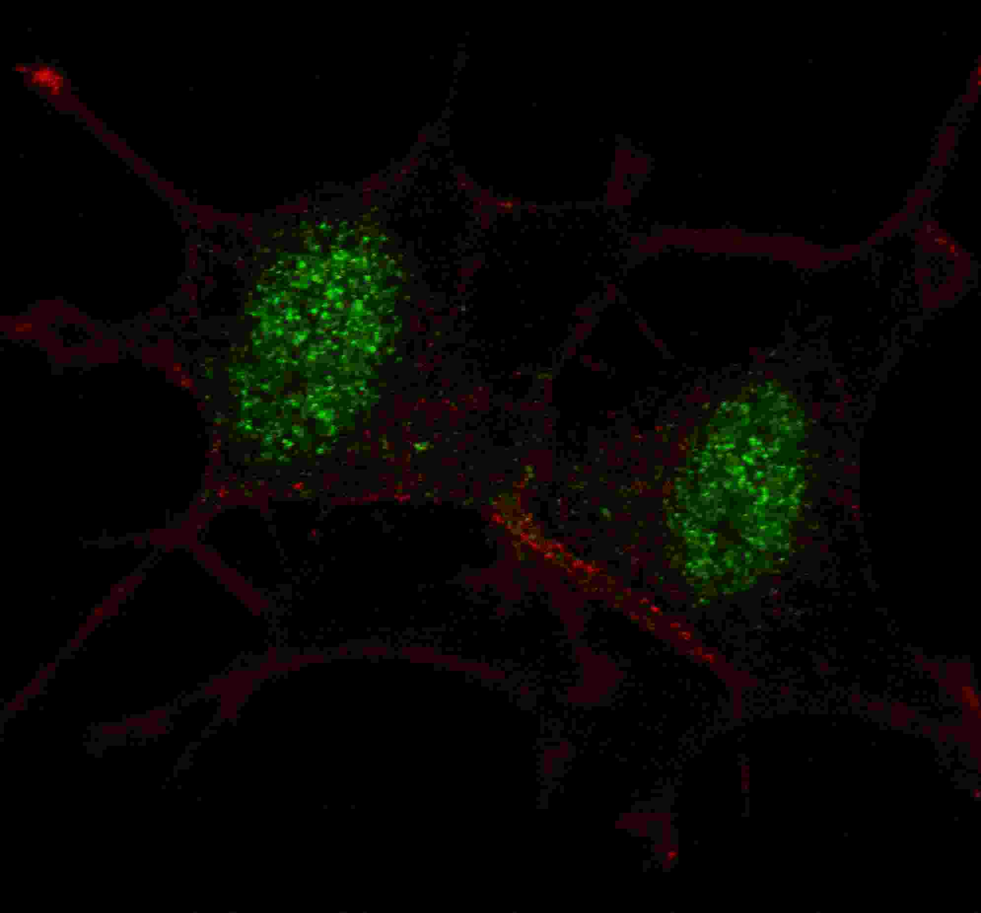
Fluorescent confocal image of SY5Y cells stained with SMAD2 (S118) antibody. SY5Y cells were fixed with 4% PFA (20 min), permeabilized with Triton X-100 (0.2%, 30 min). Cells were then incubated with AP7365a SMAD2 (S118) primary antibody (1:100, 2 h at room temperature). For secondary antibody, Alexa Fluor® 488 conjugated donkey anti-rabbit antibody (green) was used (1:1000, 1h). Nuclei were counterstained with Hoechst 33342 (blue) (10 μg/ml, 5 min). Note the highly specific localization of the SMAD2 mainly to the nucleus.Application
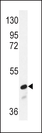
Western blot analysis of SMAD2 Antibody (S118) (Cat.# AP7365a) in NIH-3T3 cell line lysates (35ug/lane). SMAD2 (arrow) was detected using the purified Pab.Application
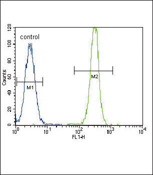
SMAD2 Antibody (S118) flow cytometric analysis of Hela cells (right histogram) compared to a negative control cell (left histogram).FITC-conjugated goat-anti-rabbit secondary antibodies were used for the analysis.| Product Name | SMAD2 Antibody (S118) |
|---|---|
| Antibody Type | Primary Antibodies |
| Antigen Alias | SMAD2; MADH2; MADR2; Mothers against decapentaplegic homolog 2; JV18-1; Mad-related protein 2; SMAD family member 2 |
| Clonality | Polyclonal |
|---|---|
| Isotype | Ig |
| Host Species | Rabbit |
| Tested Applications | IFWBFC |
| IF:1:100 WB |
|
| Species Reactivity | Human |
| Concentration | 1mg/ml |
| Gene Synonyms | MADH2 MADR2 |
|---|---|
| Alternative Names | SMAD2 MADH2 MADR2 Mothers against decapentaplegic homolog 2 JV18-1 Mad-related protein 2 SMAD family member 2 |
| Molecular Weight(MW) | 52306 Da |
| Function | Receptor-regulated SMAD (R-SMAD) that is an intracellular signal transducer and transcriptional modulator activated by TGF-beta (transforming growth factor) and activin type 1 receptor kinases. Binds the TRE element in the promoter region of many genes that are regulated by TGF-beta and, on formation of the SMAD2/SMAD4 complex, activates transcription. May act as a tumor suppressor in colorectal carcinoma. Positively regulates PDPK1 kinase activity by stimulating its dissociation from the 14-3-3 protein YWHAQ which acts as a negative regulator |
| Tissue Specificity | Expressed at high levels in skeletal muscle, heart and placenta |
| Cellular Localization | Cytoplasm. Nucleus. Note=Cytoplasmic and nuclear in the absence of TGF-beta. On TGF-beta stimulation, migrates to the nucleus when complexed with SMAD4. On dephosphorylation by phosphatase PPM1A, released from the SMAD2/SMAD4 complex, and exported out of the nucleus by interaction with RANBP1 |
| Entrez Gene | 4087 |
|---|
Application
Fluorescent confocal image of SY5Y cells stained with SMAD2 (S118) antibody. SY5Y cells were fixed with 4% PFA (20 min), permeabilized with Triton X-100 (0.2%, 30 min). Cells were then incubated with AP7365a SMAD2 (S118) primary antibody (1:100, 2 h at room temperature). For secondary antibody, Alexa Fluor® 488 conjugated donkey anti-rabbit antibody (green) was used (1:1000, 1h). Nuclei were counterstained with Hoechst 33342 (blue) (10 μg/ml, 5 min). Note the highly specific localization of the SMAD2 mainly to the nucleus.
Application
Western blot analysis of SMAD2 Antibody (S118) (Cat.# AP7365a) in NIH-3T3 cell line lysates (35ug/lane). SMAD2 (arrow) was detected using the purified Pab.
Application
SMAD2 Antibody (S118) flow cytometric analysis of Hela cells (right histogram) compared to a negative control cell (left histogram).FITC-conjugated goat-anti-rabbit secondary antibodies were used for the analysis.| Application Notes | IF:1:100 WB |
|---|
| Form | Liquid |
|---|---|
| Storage Instructions | For short-term storage, store at 4° C. For long-term storage, aliquot and store at -20ºC or below. Avoid multiple freeze-thaw cycles. |
| Storage Buffer | Purified polyclonal antibody supplied in PBS with 0.09% (W/V) sodium azide. This antibody is purified through a protein A column, followed by peptide affinity purification. |


 Data sheet for OM221448