

Application
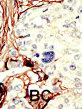
Formalin-fixed and paraffin-embedded human cancer tissue reacted with the primary antibody, which was peroxidase-conjugated to the secondary antibody, followed by DAB staining. This data demonstrates the use of this antibody for immunohistochemistry; clinical relevance has not been evaluated. BC = breast carcinoma; HC = hepatocarcinoma.IHC
The positive staining of LKB1 protein was mainly found in the cytoplasm of GC tumor cells and paracancerous normal gastric mucosa: normal gastric mucosa, positive staining (A); high/moderate differentiated GC, positive staining (B); and low differentiated GC, negative staining (C).IHC
LKB1 protein staining in normal gastric mucosa and GC tissue: (A) normal gastric mucosa (positive); (B) GC (positive); and (C) GC (negative). p53 staining in GC tissue: (D) positive; (E) punctate positive; and (F) negative. Survivin staining in GC tissue: (G) strong staining; (H) moderate staining; and (I) negative staining (×200).| Product Name | STK11 (LKB1) Antibody (C-term) |
|---|---|
| Antibody Type | Primary Antibodies |
| Clonality | Polyclonal |
|---|---|
| Isotype | Ig |
| Host Species | Rabbit |
| Tested Applications | IHC |
| IHC:1:50~100 |
|
| Species Reactivity | Human |
| Concentration | 1mg/ml |
| Gene Synonyms | LKB1 PJS |
|---|---|
| Alternative Names | STK11 LKB1 PJS Serine/threonine-protein kinase STK11 Liver kinase B1 Renal carcinoma antigen NY-REN-19 |
| Molecular Weight(MW) | 48636 Da |
| Function | Tumor suppressor serine/threonine-protein kinase that controls the activity of AMP-activated protein kinase (AMPK) family members, thereby playing a role in various processes such as cell metabolism, cell polarity, apoptosis and DNA damage response. Acts by phosphorylating the T-loop of AMPK family proteins, leading to promote their activity: phosphorylates PRKAA1, PRKAA2, BRSK1, BRSK2, MARK1, MARK2, MARK3, MARK4, NUAK1, NUAK2, SIK1, SIK2, SIK3 and SNRK but not MELK. Also phosphorylates non-AMPK family proteins such as STRADA and possibly p53/TP53 Acts as a key upstream regulator of AMPK by mediating phosphorylation and activation of AMPK catalytic subunits PRKAA1 and PRKAA2: it thereby regulates inhibition of signaling pathways that promote cell growth and proliferation when energy levels are low, glucose homeostasis in liver, activation of autophagy when cells undergo nutrient deprivation, B-cell differentiation in the germinal center in response to DNA damage. Also acts as a regulator of cellular polarity by remodeling the actin cytoskeleton. Required for cortical neurons polarization by mediating phosphorylation and activation of BRSK1 and BRSK2, leading to axon initiation and specification. Involved in DNA damage response: interacts with p53/TP53 and recruited to the CDKN1A/WAF1 promoter to participate in transcription activation Able to phosphorylate p53/TP53; the relevance of such result in vivo is however unclear and phosphorylation may be indirect and mediated by downstream STK11/LKB1 kinase NUAK1 Also acts as a mediator p53/TP53-dependent apoptosis via interaction with p53/TP53: translocates to mitochondrion during apoptosis and regulates p53/TP53-dependent apoptosis pathways |
| Tissue Specificity | Ubiquitously expressed. Strongest expression in testis and fetal liver |
| Cellular Localization | Nucleus. Cytoplasm. Membrane (By similarity). Mitochondrion. Note=A small fraction localizes at membranes (By similarity). Relocates to the cytoplasm when bound to STRAD (STRADA or STRADB) and CAB39/MO25 (CAB39/MO25alpha or CAB39L/MO25beta). Translocates to mitochondrion during apoptosis |
| Entrez Gene | 6794 |
|---|
Application
Formalin-fixed and paraffin-embedded human cancer tissue reacted with the primary antibody, which was peroxidase-conjugated to the secondary antibody, followed by DAB staining. This data demonstrates the use of this antibody for immunohistochemistry; clinical relevance has not been evaluated. BC = breast carcinoma; HC = hepatocarcinoma.
IHC
The positive staining of LKB1 protein was mainly found in the cytoplasm of GC tumor cells and paracancerous normal gastric mucosa: normal gastric mucosa, positive staining (A); high/moderate differentiated GC, positive staining (B); and low differentiated GC, negative staining (C).
IHC
LKB1 protein staining in normal gastric mucosa and GC tissue: (A) normal gastric mucosa (positive); (B) GC (positive); and (C) GC (negative). p53 staining in GC tissue: (D) positive; (E) punctate positive; and (F) negative. Survivin staining in GC tissue: (G) strong staining; (H) moderate staining; and (I) negative staining (×200).| Application Notes | IHC:1:50~100 |
|---|
| Form | Liquid |
|---|---|
| Storage Instructions | For short-term storage, store at 4° C. For long-term storage, aliquot and store at -20ºC or below. Avoid multiple freeze-thaw cycles. |
| Storage Buffer | Purified polyclonal antibody supplied in PBS with 0.09% (W/V) sodium azide. This antibody is purified through a protein G column, eluted with high and low pH buffers and neutralized immediately, followed by dialysis against PBS. |


