Application
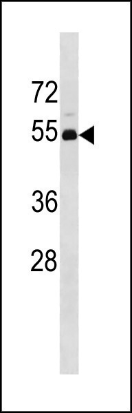
TP53 Antibody (N-term) western blot analysis in 293 cell line lysates (35ug/lane).This demonstrates the TP53 antibody detected the TP53 protein (arrow).| Product Name | TP53 Antibody (N-term) |
|---|---|
| Antibody Type | Primary Antibodies |
| Antigen Alias | TP53;P53; Cellular tumor antigen p53; Cellular tumor antigen p53; Antigen NY-CO-13; Cellular tumor antigen p53; Phosphoprotein p53; Cellular tumor antigen p53; Tumor suppressor p53 |
| Clonality | Polyclonal |
|---|---|
| Isotype | Ig |
| Host Species | Rabbit |
| Tested Applications | WB |
| WB:1:100~500 |
|
| Species Reactivity | Human |
| Concentration | 1mg/ml |
| Gene Synonyms | P53 |
|---|---|
| Alternative Names | TP53 P53 Cellular tumor antigen p53 Cellular tumor antigen p53 Antigen NY-CO-13 Cellular tumor antigen p53 Phosphoprotein p53 Cellular tumor antigen p53 Tumor suppressor p53 |
| Molecular Weight(MW) | 43653 Da |
| Function | Acts as a tumor suppressor in many tumor types; induces growth arrest or apoptosis depending on the physiological circumstances and cell type. Involved in cell cycle regulation as a trans-activator that acts to negatively regulate cell division by controlling a set of genes required for this process. One of the activated genes is an inhibitor of cyclin-dependent kinases Apoptosis induction seems to be mediated either by stimulation of BAX and FAS antigen expression, or by repression of Bcl-2 expression. In cooperation with mitochondrial PPIF is involved in activating oxidative stress-induced necrosis; te function is largely independent of transcription. Induces the transcription of long intergenic non-coding RNA p21 (lincRNA-p21) and lincRNA- Mkln1. LincRNA-p21 participates in TP53-dependent transcriptional repression leading to apoptosis and seem to have to effect on cell-cycle regulation. Implicated in Notch signaling cross-over Prevents CDK7 kinase activity when associated to CAK complex in response to DNA damage, thus stopping cell cycle progression Isoform 2 enhances the transactivation activity of isoform 1 from some but not all TP53-inducible promoters. Isoform 4 suppresses transactivation activity and impairs growth suppression mediated by isoform 1. Isoform 7 inhibits isoform 1-mediated apoptosis |
| Tissue Specificity | Ubiquitous. Isoforms are expressed in a wide range of normal tissues but in a tissue-dependent manner. Isoform 2 is expressed in most normal tissues but is not detected in brain, lung, prostate, muscle, fetal brain, spinal cord and fetal liver. Isoform 3 is expressed in most normal tissues but is not detected in lung, spleen, testis, fetal brain, spinal cord and fetal liver. Isoform 7 is expressed in most normal tissues but is not detected in prostate, uterus, skeletal muscle and breast Isoform 8 is detected only in colon, bone marrow, testis, fetal brain and intestine. Isoform 9 is expressed in most normal tissues but is not detected in brain, heart, lung, fetal liver, salivary gland, breast or intestine |
| Cellular Localization | Cytoplasm. Nucleus. Nucleus, PML body. Endoplasmic reticulum. Mitochondrion matrix. Note=Interaction with BANP promotes nuclear localization. Recruited into PML bodies together with CHEK2. Translocates to mitochondria upon oxidative stress Isoform 2: Nucleus. Cytoplasm. Note=Localized mainly in the nucleus with minor staining in the cytoplasm Isoform 4: Nucleus. Cytoplasm. Note=Predominantly nuclear but translocates to the cytoplasm following cell stress Isoform 8: Nucleus. Cytoplasm. Note=Localized in both nucleus and cytoplasm in most cells. In some cells, forms foci in the nucleus that are different from nucleoli |
| Entrez Gene | 7157 |
|---|
Application
TP53 Antibody (N-term) western blot analysis in 293 cell line lysates (35ug/lane).This demonstrates the TP53 antibody detected the TP53 protein (arrow).| Application Notes | WB:1:100~500 |
|---|
| Form | Liquid |
|---|---|
| Storage Instructions | For short-term storage, store at 4° C. For long-term storage, aliquot and store at -20ºC or below. Avoid multiple freeze-thaw cycles. |
| Storage Buffer | Purified polyclonal antibody supplied in PBS with 0.09% (W/V) sodium azide. This antibody is purified through a protein A column, followed by peptide affinity purification. |


