Application
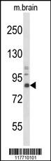
Western blot analysis of TrkA Antibody in mouse brain tissue lysates (35ug/lane). TrkA (arrow) was detected using the purified Pab.Application
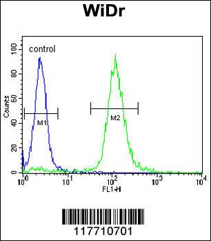
TrkA Antibody flow cytometric analysis of WiDr cells (right histogram) compared to a negative control cell (left histogram).FITC-conjugated goat-anti-rabbit secondary antibodies were used for the analysis.Application
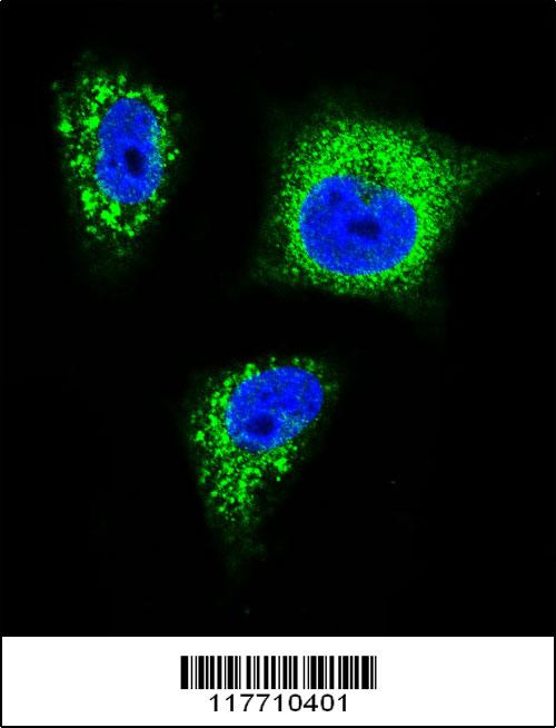
Confocal immunofluorescent analysis of TrkA Antibody(Cat#AP7686f) with MDA-MB231 cell followed by Alexa Fluor 488-conjugated goat anti-rabbit lgG (green).DAPI was used to stain the cell nuclear (blue).| Product Name | TrkA Antibody |
|---|---|
| Antibody Type | Primary Antibodies |
| Antigen Alias | NTRK1; MTC; TRK; TRKA; High affinity nerve growth factor receptor; Neurotrophic tyrosine kinase receptor type 1; TRK1-transforming tyrosine kinase protein; Tropomyosin-related kinase A; Tyrosine kinase receptor; Tyrosine kinase receptor A; gp140trk; p140-T |
| Clonality | Polyclonal |
|---|---|
| Isotype | Ig |
| Host Species | Rabbit |
| Tested Applications | WBFCIF |
| WB:1:50~100 FC |
|
| Species Reactivity | Human |
| Concentration | 1mg/ml |
| Gene Synonyms | MTC TRK TRKA |
|---|---|
| Alternative Names | NTRK1 MTC TRK TRKA High affinity nerve growth factor receptor Neurotrophic tyrosine kinase receptor type 1 TRK1-transforming tyrosine kinase protein Tropomyosin-related kinase A Tyrosine kinase receptor Tyrosine kinase receptor A gp140trk p140-T |
| Molecular Weight(MW) | 87497 Da |
| Function | Receptor tyrosine kinase involved in the development and the maturation of the central and peripheral nervous systems through regulation of proliferation, differentiation and survival of sympathetic and nervous neurons. High affinity receptor for NGF which is its primary ligand, it can also bind and be activated by NTF3/neurotrophin-3. However, NTF3 only supports axonal extension through NTRK1 but has no effect on neuron survival. Upon dimeric NGF ligand-binding, undergoes homodimerization, autophosphorylation and activation. Recruits, phosphorylates and/or activates several downstream effectors including SHC1, FRS2, SH2B1, SH2B2 and PLCG1 that regulate distinct overlapping signaling cascades driving cell survival and differentiation Through SHC1 and FRS2 activates a GRB2-Ras-MAPK cascade that regulates cell differentiation and survival. Through PLCG1 controls NF-Kappa-B activation and the transcription of genes involved in cell survival. Through SHC1 and SH2B1 controls a Ras- PI3 kinase-AKT1 signaling cascade that is also regulating survival. In absence of ligand and activation, may promote cell death, making the survival of neurons dependent on trophic factors |
| Tissue Specificity | Isoform TrkA-I is found in most non-neuronal tissues. Isoform TrkA-II is primarily expressed in neuronal cells TrkA-III is specifically expressed by pluripotent neural stem and neural crest progenitors |
| Cellular Localization | Cell membrane; Single-pass type I membrane protein. Early endosome membrane; Single-pass type I membrane protein (By similarity). Note=Internalized to endosomes upon binding of NGF or NTF3 and further transported to the cell body via a retrograde axonal transport (By similarity) |
| Entrez Gene | 4914 |
|---|
Application
Western blot analysis of TrkA Antibody in mouse brain tissue lysates (35ug/lane). TrkA (arrow) was detected using the purified Pab.
Application
TrkA Antibody flow cytometric analysis of WiDr cells (right histogram) compared to a negative control cell (left histogram).FITC-conjugated goat-anti-rabbit secondary antibodies were used for the analysis.
Application
Confocal immunofluorescent analysis of TrkA Antibody(Cat#AP7686f) with MDA-MB231 cell followed by Alexa Fluor 488-conjugated goat anti-rabbit lgG (green).DAPI was used to stain the cell nuclear (blue).| Application Notes | WB:1:50~100 FC |
|---|
| Form | Liquid |
|---|---|
| Storage Instructions | For short-term storage, store at 4° C. For long-term storage, aliquot and store at -20ºC or below. Avoid multiple freeze-thaw cycles. |
| Storage Buffer | Purified polyclonal antibody supplied in PBS with 0.09% (W/V) sodium azide. This antibody is prepared by Saturated Ammonium Sulfate (SAS) precipitation followed by dialysis against PBS. |


