Application
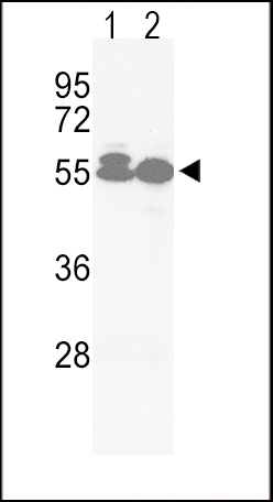
Western blot analysis of hACVRL1-L489 in Jurkat(lane 1), HepG2(lane 2) cell line lysates (35ug/lane). ACVRL1 (arrow) was detected using the purified Pab.Application
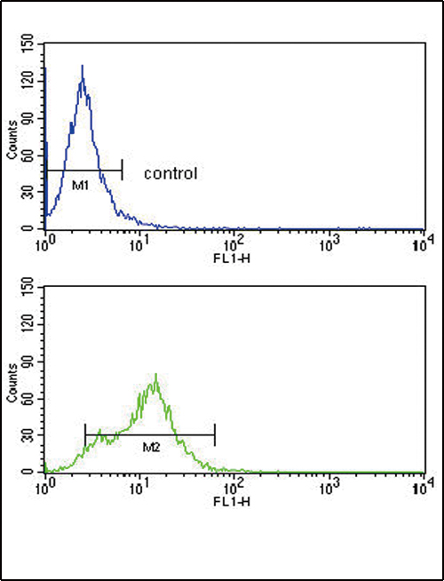
ACVRL1 Antibody (C-term) flow cytometry analysis of HepG2 cells (bottom histogram) compared to a negative control cell (top histogram). FITC-conjugated goat-anti-rabbit secondary antibodies were used for the analysis.| Product Name | ACVRL1 Antibody (C-term) |
|---|---|
| Antibody Type | Primary Antibodies |
| Antigen Alias | ACVRL1; ACVRLK1; ALK1; Serine/threonine-protein kinase receptor R3; Activin receptor-like kinase 1; TGF-B superfamily receptor type I |
| Clonality | Polyclonal |
|---|---|
| Isotype | Ig |
| Host Species | Rabbit |
| Tested Applications | WBFC |
| WB:1:100~500 FC |
|
| Species Reactivity | Human |
| Concentration | 1mg/ml |
| Gene Synonyms | ACVRLK1 ALK1 |
|---|---|
| Alternative Names | ACVRL1 ACVRLK1 ALK1 Serine/threonine-protein kinase receptor R3 Activin receptor-like kinase 1 TGF-B superfamily receptor type I |
| Molecular Weight(MW) | 56124 Da |
| Function | Type I receptor for TGF-beta family ligands BMP9/GDF2 and BMP10 and important regulator of normal blood vessel development. On ligand binding, forms a receptor complex consisting of two type II and two type I transmembrane serine/threonine kinases. Type II receptors phosphorylate and activate type I receptors which autophosphorylate, then bind and activate SMAD transcriptional regulators. May bind activin as well |
| Tissue Specificity | This ACVRL1 antibody is generated from rabbits immunized with a KLH conjugated synthetic peptide between 474~503 amino acids from the C-terminal region of human ACVRL1. |
| Cellular Localization | Membrane; Single-pass type I membrane protein |
| Entrez Gene | 94 |
|---|
Application
Western blot analysis of hACVRL1-L489 in Jurkat(lane 1), HepG2(lane 2) cell line lysates (35ug/lane). ACVRL1 (arrow) was detected using the purified Pab.
Application
ACVRL1 Antibody (C-term) flow cytometry analysis of HepG2 cells (bottom histogram) compared to a negative control cell (top histogram). FITC-conjugated goat-anti-rabbit secondary antibodies were used for the analysis.| Application Notes | WB:1:100~500 FC |
|---|
| Form | Liquid |
|---|---|
| Storage Instructions | For short-term storage, store at 4° C. For long-term storage, aliquot and store at -20ºC or below. Avoid multiple freeze-thaw cycles. |
| Storage Buffer | Purified polyclonal antibody supplied in PBS with 0.09% (W/V) sodium azide. This antibody is purified through a protein G column, eluted with high and low pH buffers and neutralized immediately, followed by dialysis against PBS. |


