Application
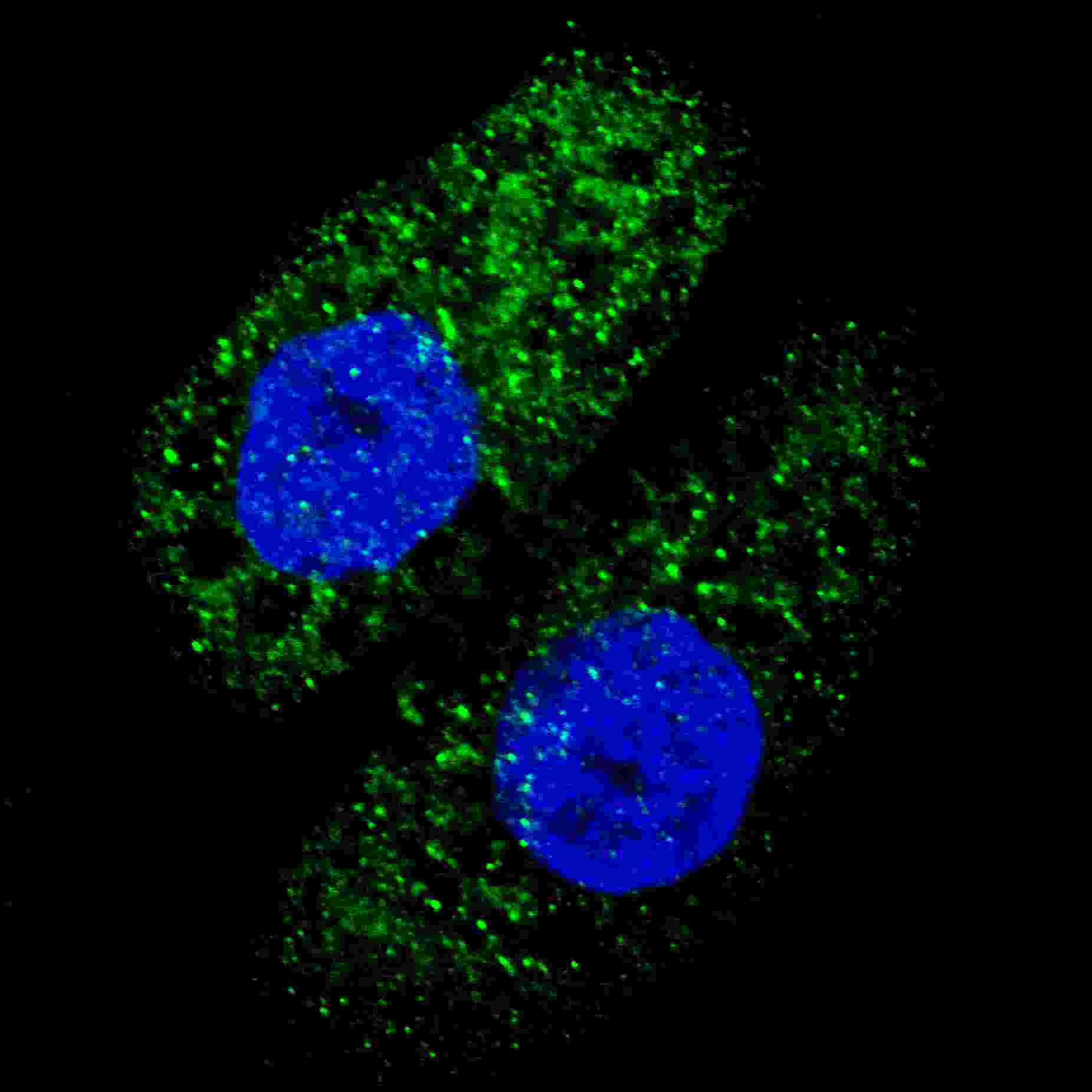
Fluorescent image of U251 cells stained with APG5 antibody. U251 cells were treated with Chloroquine (50 μM,16h), then fixed with 4% PFA (20 min), permeabilized with Triton X-100 (0.2%, 30 min). Cells were then incubated with AP1812c APG5 primary antibody (1:200, 2 h at room temperature). For secondary antibody, Alexa Fluor® 488 conjugated donkey anti-rabbit antibody (green) was used (1:1000, 1h). Nuclei were counterstained with Hoechst 33342 (blue) (10 μg/ml, 5 min). ATG5 immunoreactivity is localized to autophagic vacuoles in the cytoplasm of U251 cells.Application
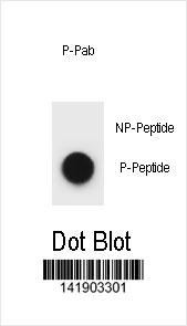
APG5 Antibody western blot analysis in UM cell line lysates (35ug/lane).This demonstrates the APG5 antibody detected the APG5 protein (arrow).Application
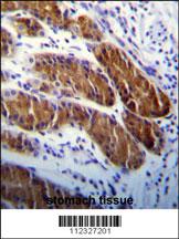
APG5 Antibody immunohistochemistry analysis in formalin fixed and paraffin embedded human stomach tissue followed by peroxidase conjugation of the secondary antibody and DAB staining.This data demonstrates the use of APG5 Antibody for immunohistochemistry. Clinical relevance has not been evaluated.| Product Name | APG5 Antibody |
|---|---|
| Antibody Type | Primary Antibodies |
| Antigen Alias | ATG5; APG5L; ASP; Autophagy protein 5; APG5-like; Apoptosis-specific protein |
| Clonality | Polyclonal |
|---|---|
| Isotype | Ig |
| Host Species | Rabbit |
| Tested Applications | IFWBIHC |
| IF:1:200 WB |
|
| Species Reactivity | Human |
| Concentration | 1mg/ml |
| Gene Synonyms | APG5L ASP |
|---|---|
| Alternative Names | ATG5 APG5L ASP Autophagy protein 5 APG5-like Apoptosis-specific protein |
| Molecular Weight(MW) | 32447 Da |
| Function | Required for autophagy. Conjugates to ATG12 and associates with isolation membrane to form cup-shaped isolation membrane and autophagosome. The conjugate detaches from the membrane immediately before or after autophagosome formation is completed (By similarity) |
| Tissue Specificity | Ubiquitous. The mRNA is present at similar levels in viable and apoptotic cells, whereas the protein is dramatically highly expressed in apoptotic cells |
| Cellular Localization | Cytoplasm. Note=Colocalizes with nonmuscle actin |
| Entrez Gene | 9474 |
|---|
Application
Fluorescent image of U251 cells stained with APG5 antibody. U251 cells were treated with Chloroquine (50 μM,16h), then fixed with 4% PFA (20 min), permeabilized with Triton X-100 (0.2%, 30 min). Cells were then incubated with AP1812c APG5 primary antibody (1:200, 2 h at room temperature). For secondary antibody, Alexa Fluor® 488 conjugated donkey anti-rabbit antibody (green) was used (1:1000, 1h). Nuclei were counterstained with Hoechst 33342 (blue) (10 μg/ml, 5 min). ATG5 immunoreactivity is localized to autophagic vacuoles in the cytoplasm of U251 cells.
Application
APG5 Antibody western blot analysis in UM cell line lysates (35ug/lane).This demonstrates the APG5 antibody detected the APG5 protein (arrow).
Application
APG5 Antibody immunohistochemistry analysis in formalin fixed and paraffin embedded human stomach tissue followed by peroxidase conjugation of the secondary antibody and DAB staining.This data demonstrates the use of APG5 Antibody for immunohistochemistry. Clinical relevance has not been evaluated.| Application Notes | IF:1:200 WB |
|---|
| Form | Liquid |
|---|---|
| Storage Instructions | For short-term storage, store at 4° C. For long-term storage, aliquot and store at -20ºC or below. Avoid multiple freeze-thaw cycles. |
| Storage Buffer | Purified polyclonal antibody supplied in PBS with 0.09% (W/V) sodium azide. This antibody is purified through a protein G column, eluted with high and low pH buffers and neutralized immediately, followed by dialysis against PBS. |


