Application
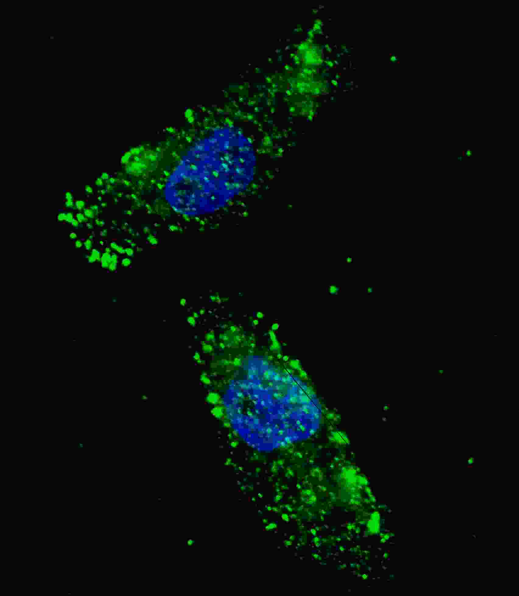
Fluorescent image of U251 cells stained with APG1 (ULK1) (Center) antibody. U251 cells were treated with Chloroquine (50 μM,16h), then fixed with 4% PFA (20 min), permeabilized with Triton X-100 (0.2%, 30 min). Cells were then incubated with AP8104b APG1 (ULK1) (Center) primary antibody (1:100, 2 h at room temperature). For secondary antibody, Alexa Fluor® 488 conjugated donkey anti-rabbit antibody (green) was used (1:1000, 1h). Nuclei were counterstained with Hoechst 33342 (blue) (10 μg/ml, 5 min). APG1 (ULK1) immunoreactivity is localized to autophagic vacuoles in the cytoplasm of U251 cells.Application
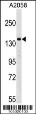
Western blot analysis of anti-ULK1 Pab in A2058 cell lysate. ULK1 (arrow) was detected using purified Pab. Secondary HRP-anti-rabbit was used for signal visualization with chemiluminescence.| Product Name | APG1 (ULK1) Antibody (Center) |
|---|---|
| Antibody Type | Primary Antibodies |
| Antigen Alias | ULK1; KIAA0722; Serine/threonine-protein kinase ULK1; Autophagy-related protein 1 homolog; Unc-51-like kinase 1 |
| Clonality | Polyclonal |
|---|---|
| Isotype | Ig |
| Host Species | Rabbit |
| Tested Applications | IFWB |
| IF:1:100 WB |
|
| Species Reactivity | Human |
| Concentration | 1mg/ml |
| Gene Synonyms | KIAA0722 |
|---|---|
| Alternative Names | ULK1 KIAA0722 Serine/threonine-protein kinase ULK1 Autophagy-related protein 1 homolog Unc-51-like kinase 1 |
| Molecular Weight(MW) | 112631 Da |
| Function | Serine/threonine-protein kinase involved in autophagy in response to starvation. Acts upstream of phosphatidylinositol 3- kinase PIK3C3 to regulate the formation of autophagophores, the precursors of autophagosomes. Part of regulatory feedback loops in autophagy: acts both as a downstream effector and negative regulator of mammalian target of rapamycin complex 1 (mTORC1) via interaction with RPTOR. Activated via phosphorylation by AMPK and also acts as a regulator of AMPK by mediating phosphorylation of AMPK subunits PRKAA1, PRKAB2 and PRKAG1, leading to negatively regulate AMPK activity. May phosphorylate ATG13/KIAA0652 and RPTOR; however such data need additional evidences. Plays a role early in neuronal differentiation and is required for granule cell axon formation |
| Tissue Specificity | Ubiquitously expressed. Detected in the following adult tissues: skeletal muscle, heart, pancreas, brain, placenta, liver, kidney, and lung |
| Cellular Localization | Cytoplasm, cytosol (By similarity). Preautophagosomal structure (By similarity). Note=Under starvation conditions, is localized to puncate structures primarily representing the isolation membrane that sequesters a portion of the cytoplasm resulting in the formation of an autophagosome (By similarity) |
| Entrez Gene | 8408 |
|---|
Application
Fluorescent image of U251 cells stained with APG1 (ULK1) (Center) antibody. U251 cells were treated with Chloroquine (50 μM,16h), then fixed with 4% PFA (20 min), permeabilized with Triton X-100 (0.2%, 30 min). Cells were then incubated with AP8104b APG1 (ULK1) (Center) primary antibody (1:100, 2 h at room temperature). For secondary antibody, Alexa Fluor® 488 conjugated donkey anti-rabbit antibody (green) was used (1:1000, 1h). Nuclei were counterstained with Hoechst 33342 (blue) (10 μg/ml, 5 min). APG1 (ULK1) immunoreactivity is localized to autophagic vacuoles in the cytoplasm of U251 cells.
Application
Western blot analysis of anti-ULK1 Pab in A2058 cell lysate. ULK1 (arrow) was detected using purified Pab. Secondary HRP-anti-rabbit was used for signal visualization with chemiluminescence.| Application Notes | IF:1:100 WB |
|---|
| Form | Liquid |
|---|---|
| Storage Instructions | For short-term storage, store at 4° C. For long-term storage, aliquot and store at -20ºC or below. Avoid multiple freeze-thaw cycles. |
| Storage Buffer | Purified polyclonal antibody supplied in PBS with 0.09% (W/V) sodium azide. This antibody is purified through a protein G column, eluted with high and low pH buffers and neutralized immediately, followed by dialysis against PBS. |


