Application
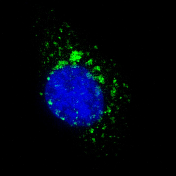
Fluorescent image of U251 cells stained with ATG4B antibody. U251 cells were treated with Chloroquine (50 μM,16h), then fixed with 4% PFA (20 min), permeabilized with Triton X-100 (0.2%, 30 min). Cells were then incubated with AP1809f ATG4B primary antibody (1:100, 2 h at room temperature). For secondary antibody, Alexa Fluor® 488 conjugated donkey anti-rabbit antibody (green) was used (1:1000, 1h). Nuclei were counterstained with Hoechst 33342 (blue) (10 μg/ml, 5 min). ATG4B immunoreactivity is localized to autophagic vacuoles in the cytoplasm of U251 cells.Application
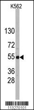
Western blot analysis of anti-APG4B Antibody (S309) in K562 cell line lysates (35ug/lane). APG4B (arrow) was detected using the purified Pab (1:60 dilution).Application
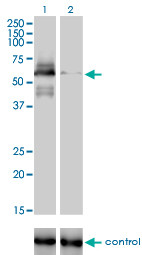
Western blot analysis of APG4B (arrow) using rabbit polyclonal APG4B Antibody (S309) . 293 cell lysates (2 ug/lane) either nontransfected (Lane 1) or transiently transfected with the APG4B gene (Lane 2) (Origene Technologies).Application
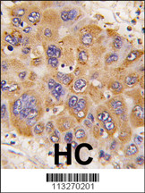
Formalin-fixed and paraffin-embedded human hepatocarcinoma tissue reacted with Phospho-APG4B-pS309, which was peroxidase-conjugated to the secondary antibody, followed by DAB staining. This data demonstrates the use of this antibody for immunohistochemistry; clinical relevance has not been evaluated.| Product Name | ATG4B Antibody |
|---|---|
| Antibody Type | Primary Antibodies |
| Clonality | Polyclonal |
|---|---|
| Isotype | Ig |
| Host Species | Rabbit |
| Tested Applications | IFWBIHC |
| IF:1:100 WB |
|
| Species Reactivity | Human |
| Concentration | 1mg/ml |
| Gene Synonyms | APG4B, AUTL1, KIAA0943 |
|---|---|
| Alternative Names | ATG4B APG4B AUTL1 KIAA0943 Cysteine protease ATG4B AUT-like 1 cysteine endopeptidase Autophagin-1 Autophagy-related cysteine endopeptidase 1 Autophagy-related protein 4 homolog B |
| Molecular Weight(MW) | 44294 Da |
| Function | Cysteine protease required for autophagy, which cleaves the C-terminal part of MAP1LC3, GABARAPL1, GABARAPL2 and GABARAP, allowing the liberation of form I. A subpopulation of form I is subsequently converted to a smaller form (form II). Form II, with a revealed C-terminal glycine, is considered to be the phosphatidylethanolamine (PE)-conjugated form, and has the capacity for the binding to autophagosomes. Also mediates the lipid deconjugation required for target recycling |
| Tissue Specificity | Mainly expressed in the skeletal muscle, followed by brain, heart, liver and pancreas |
| Cellular Localization | Cytoplasm (Probable). |
| Entrez Gene | 23192 |
|---|
Application
Fluorescent image of U251 cells stained with ATG4B antibody. U251 cells were treated with Chloroquine (50 μM,16h), then fixed with 4% PFA (20 min), permeabilized with Triton X-100 (0.2%, 30 min). Cells were then incubated with AP1809f ATG4B primary antibody (1:100, 2 h at room temperature). For secondary antibody, Alexa Fluor® 488 conjugated donkey anti-rabbit antibody (green) was used (1:1000, 1h). Nuclei were counterstained with Hoechst 33342 (blue) (10 μg/ml, 5 min). ATG4B immunoreactivity is localized to autophagic vacuoles in the cytoplasm of U251 cells.
Application
Western blot analysis of anti-APG4B Antibody (S309) in K562 cell line lysates (35ug/lane). APG4B (arrow) was detected using the purified Pab (1:60 dilution).
Application
Western blot analysis of APG4B (arrow) using rabbit polyclonal APG4B Antibody (S309) . 293 cell lysates (2 ug/lane) either nontransfected (Lane 1) or transiently transfected with the APG4B gene (Lane 2) (Origene Technologies).
Application
Formalin-fixed and paraffin-embedded human hepatocarcinoma tissue reacted with Phospho-APG4B-pS309, which was peroxidase-conjugated to the secondary antibody, followed by DAB staining. This data demonstrates the use of this antibody for immunohistochemistry; clinical relevance has not been evaluated.| Application Notes | IF:1:100 WB |
|---|
| Form | Liquid |
|---|---|
| Storage Instructions | For short-term storage, store at 4° C. For long-term storage, aliquot and store at -20ºC or below. Avoid multiple freeze-thaw cycles. |
| Storage Buffer | Purified polyclonal antibody supplied in PBS with 0.09% (W/V) sodium azide. This antibody is purified through a protein A column, followed by peptide affinity purification. |


