Application
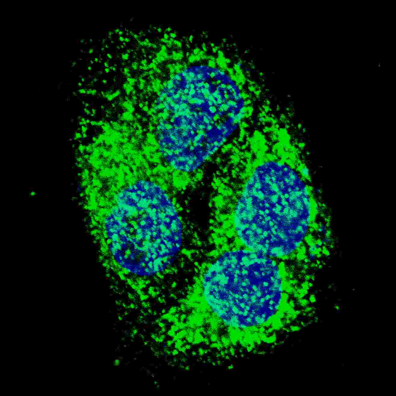
Fluorescent confocal image of HepG2 cells stained with BNIP3 (BH3 Domain Specific) antibody. HepG2 cells were fixed with 4% PFA (20 min), permeabilized with Triton X-100 (0.2%, 30 min). Cells were then incubated with AP1321a BNIP3 (BH3 Domain Specific) primary antibody (1:500, 2 h at room temperature). For secondary antibody, Alexa Fluor® 488 conjugated donkey anti-rabbit antibody (green) was used (1:1000, 1h). Nuclei were counterstained with Hoechst 33342 (blue) (10 μg/ml, 5 min). BNIP3 immunoreactivity is localized to the cytoplasm of HepG2 cells.Application
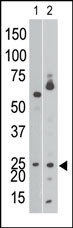
The anti-NIP3 BH3 domain Pab is used in Western blot to detect NIP3 BH3 in Ramos cell lysate (lane 1) and in mouse brain tissue lysate (lane 2).Application
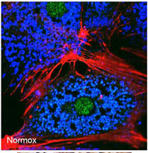
Freshly isolated mouse hepatocytes plated on coverslips (2 x10⁵ cells/22-mm glass coverslip) were cultured under normoxic conditions for 6 hr. The cells were then fixed in 2% paraformaldehyde in PBS for 1 hr, and processed for confocal immunofluorescence (red: F-actin, blue: ATP-synthase, green: BNIP3). Fluorescence labeling of BNIP3 accomplished with anti-BNIP3 antibody Cat # AP1321a. Data courtesy of Ruben Zamora, University of Pittsburgh.Application
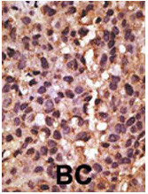
Formalin-fixed and paraffin-embedded human cancer tissue reacted with the primary antibody, which was peroxidase-conjugated to the secondary antibody, followed by DAB staining. This data demonstrates the use of this antibody for immunohistochemistry; clinical relevance has not been evaluated. BC = breast carcinoma; HC = hepatocarcinoma.| Product Name | BNIP3 Antibody (BH3 Domain Specific) |
|---|---|
| Antibody Type | Primary Antibodies |
| Antigen Alias | BNIP3; NIP3; BCL2/adenovirus E1B 19 kDa protein-interacting protein 3 |
| Clonality | Polyclonal |
|---|---|
| Isotype | Ig |
| Host Species | Rabbit |
| Tested Applications | IFWBIHC |
| IF:1:50~100 WB |
|
| Species Reactivity | Human |
| Concentration | 1mg/ml |
| Gene Synonyms | NIP3 |
|---|---|
| Alternative Names | BNIP3 NIP3 BCL2/adenovirus E1B 19 kDa protein-interacting protein 3 |
| Molecular Weight(MW) | 21541 Da |
| Function | Apoptosis-inducing protein that can overcome BCL2 suppression. May play a role in repartitioning calcium between the two major intracellular calcium stores in association with BCL2 Involved in mitochondrial quality control via its interaction with SPATA18/MIEAP: in response to mitochondrial damage, participates to mitochondrial protein catabolic process (also named MALM) leading to the degradation of damaged proteins inside mitochondria. The physical interaction of SPATA18/MIEAP, BNIP3 and BNIP3L/NIX at the mitochondrial outer membrane regulates the opening of a pore in the mitochondrial double membrane in order to mediate the translocation of lysosomal proteins from the cytoplasm to the mitochondrial matrix. Plays an important role in the calprotectin (S100A8/A9)-induced cell death pathway |
| Tissue Specificity | This BNIP3 Antibody antibody is generated from rabbits immunized with a KLH conjugated synthetic peptide selected from the N-terminal region (BH3 domain) of human NIP3. |
| Cellular Localization | Mitochondrion. Mitochondrion outer membrane; Single-pass membrane protein. Note=Coexpression with the EIB 19- kDa protein results in a shift in NIP3 localization pattern to the nuclear envelope. Colocalizes with ACAA2 in the mitochondria Colocalizes with SPATA18 at the mitochondrion outer membrane |
| Entrez Gene | 664 |
|---|
Application
Fluorescent confocal image of HepG2 cells stained with BNIP3 (BH3 Domain Specific) antibody. HepG2 cells were fixed with 4% PFA (20 min), permeabilized with Triton X-100 (0.2%, 30 min). Cells were then incubated with AP1321a BNIP3 (BH3 Domain Specific) primary antibody (1:500, 2 h at room temperature). For secondary antibody, Alexa Fluor® 488 conjugated donkey anti-rabbit antibody (green) was used (1:1000, 1h). Nuclei were counterstained with Hoechst 33342 (blue) (10 μg/ml, 5 min). BNIP3 immunoreactivity is localized to the cytoplasm of HepG2 cells.
Application
The anti-NIP3 BH3 domain Pab is used in Western blot to detect NIP3 BH3 in Ramos cell lysate (lane 1) and in mouse brain tissue lysate (lane 2).
Application
Freshly isolated mouse hepatocytes plated on coverslips (2 x10⁵ cells/22-mm glass coverslip) were cultured under normoxic conditions for 6 hr. The cells were then fixed in 2% paraformaldehyde in PBS for 1 hr, and processed for confocal immunofluorescence (red: F-actin, blue: ATP-synthase, green: BNIP3). Fluorescence labeling of BNIP3 accomplished with anti-BNIP3 antibody Cat # AP1321a. Data courtesy of Ruben Zamora, University of Pittsburgh.
Application
Formalin-fixed and paraffin-embedded human cancer tissue reacted with the primary antibody, which was peroxidase-conjugated to the secondary antibody, followed by DAB staining. This data demonstrates the use of this antibody for immunohistochemistry; clinical relevance has not been evaluated. BC = breast carcinoma; HC = hepatocarcinoma.| Application Notes | IF:1:50~100 WB |
|---|
| Form | Liquid |
|---|---|
| Storage Instructions | For short-term storage, store at 4° C. For long-term storage, aliquot and store at -20ºC or below. Avoid multiple freeze-thaw cycles. |
| Storage Buffer | Purified polyclonal antibody supplied in PBS with 0.09% (W/V) sodium azide. This antibody is purified through a protein G column, eluted with high and low pH buffers and neutralized immediately, followed by dialysis against PBS. |


