Application
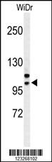
Western blot analysis of CRNKL1 Antibody (Center) in WiDr cell line lysates (35ug/lane). CRNKL1 (arrow) was detected using the purified Pab.Application
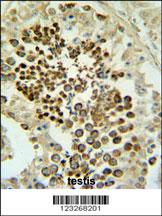
CRNKL1 Antibody (Center) IHC analysis in formalin fixed and paraffin embedded testis tissue followed by peroxidase conjugation of the secondary antibody and DAB staining. This data demonstrates the use of the CRNKL1 Antibody (Center) for immunohistochemistry. Clinical relevance has not been evaluated.Application
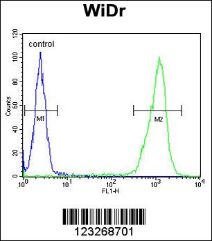
CRNKL1 Antibody (Center) flow cytometric analysis of WiDr cells (right histogram) compared to a negative control cell (left histogram).FITC-conjugated goat-anti-rabbit secondary antibodies were used for the analysis.| Product Name | CRNKL1 Antibody (Center) |
|---|---|
| Antibody Type | Primary Antibodies |
| Antigen Alias | CRNKL1; CRN; Crooked neck-like protein 1; Crooked neck homolog |
| Clonality | Polyclonal |
|---|---|
| Isotype | Ig |
| Host Species | Rabbit |
| Tested Applications | WBIHCFC |
| WB:1:100~500 IHC |
|
| Species Reactivity | Human |
| Concentration | 1mg/ml |
| Gene Synonyms | CRN |
|---|---|
| Alternative Names | CRNKL1 CRN Crooked neck-like protein 1 Crooked neck homolog |
| Molecular Weight(MW) | 100452 Da |
| Function | Involved in pre-mRNA splicing process. |
| Tissue Specificity | Widely expressed. Highly expressed in testis. Not expressed in brain and lung |
| Cellular Localization | Nucleus speckle. Cytoplasm. Note=Colocalizes with core spliceosomal snRNP proteins. Also diffusely expressed in cytoplasm |
| Entrez Gene | 51340 |
|---|
Application
Western blot analysis of CRNKL1 Antibody (Center) in WiDr cell line lysates (35ug/lane). CRNKL1 (arrow) was detected using the purified Pab.
Application
CRNKL1 Antibody (Center) IHC analysis in formalin fixed and paraffin embedded testis tissue followed by peroxidase conjugation of the secondary antibody and DAB staining. This data demonstrates the use of the CRNKL1 Antibody (Center) for immunohistochemistry. Clinical relevance has not been evaluated.
Application
CRNKL1 Antibody (Center) flow cytometric analysis of WiDr cells (right histogram) compared to a negative control cell (left histogram).FITC-conjugated goat-anti-rabbit secondary antibodies were used for the analysis.| Application Notes | WB:1:100~500 IHC |
|---|
| Form | Liquid |
|---|---|
| Storage Instructions | For short-term storage, store at 4° C. For long-term storage, aliquot and store at -20ºC or below. Avoid multiple freeze-thaw cycles. |
| Storage Buffer | Purified polyclonal antibody supplied in PBS with 0.09% (W/V) sodium azide. This antibody is purified through a protein A column, followed by peptide affinity purification. |


 Data sheet for OM230406