Application
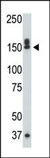
The DAPK1 polyclonal antibody is used in Western blot to detect DAPK1 in mouse liver tissue lysate.Application
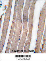
Formalin-fixed and paraffin-embedded human skeletal muscle tissue reacted with DAPK1 antibody (C-term) , which was peroxidase-conjugated to the secondary antibody, followed by DAB staining. This data demonstrates the use of this antibody for immunohistochemistry; clinical relevance has not been evaluated.Application
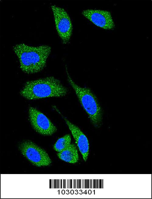
Confocal immunofluorescent analysis of DAP Kinase 1 (DAPK1) Antibody (C-term) with A549 cell followed by Alexa Fluor® 489-conjugated goat anti-rabbit lgG (green). DAPI was used to stain the cell nuclear (blue).| Product Name | DAPK1 Antibody (C-term) |
|---|---|
| Antibody Type | Primary Antibodies |
| Antigen Alias | DAPK1; DAPK; Death-associated protein kinase 1 |
| Clonality | Polyclonal |
|---|---|
| Isotype | Ig |
| Host Species | Rabbit |
| Tested Applications | WBIHCIF |
| WB:1:100~500 IHC |
|
| Species Reactivity | Human |
| Concentration | 1mg/ml |
| Gene Synonyms | DAPK |
|---|---|
| Alternative Names | DAPK1 DAPK Death-associated protein kinase 1 |
| Molecular Weight(MW) | 160046 Da |
| Function | Calcium/calmodulin-dependent serine/threonine kinase involved in multiple cellular signaling pathways that trigger cell survival, apoptosis, and autophagy. Regulates both type I apoptotic and type II autophagic cell deaths signal, depending on the cellular setting. The former is caspase-dependent, while the latter is caspase-independent and is characterized by the accumulation of autophagic vesicles. Phosphorylates PIN1 resulting in inhibition of its catalytic activity, nuclear localization, and cellular function. Phosphorylates TPM1, enhancing stress fiber formation in endothelial cells. Phosphorylates STX1A and significantly decreases its binding to STXBP1. Phosphorylates PRKD1 and regulates JNK signaling by binding and activating PRKD1 under oxidative stress. Phosphorylates BECN1, reducing its interaction with BCL2 and BCL2L1 and promoting the induction of autophagy. Phosphorylates TSC2, disrupting the TSC1-TSC2 complex and stimulating mTORC1 activity in a growth factor-dependent pathway. Phosphorylates RPS6, MYL9 and DAPK3. Acts as a signaling amplifier of NMDA receptors at extrasynaptic sites for mediating brain damage in stroke. Cerebral ischemia recruits DAPK1 into the NMDA receptor complex and it phosphorylates GRINB at Ser-1303 inducing injurious Ca(2+) influx through NMDA receptor channels, resulting in an irreversible neuronal death. Required together with DAPK3 for phosphorylation of RPL13A upon interferon-gamma activation which is causing RPL13A involvement in transcript- selective translation inhibition |
| Tissue Specificity | Isoform 2 is expressed in normal intestinal tissue as well as in colorectal carcinomas |
| Cellular Localization | Isoform 1: Cytoplasm. Cytoplasm, cytoskeleton. Note=Colocalizes with MAP1B in the microtubules and cortical actin fibers |
| Entrez Gene | 1612 |
|---|
Application
The DAPK1 polyclonal antibody is used in Western blot to detect DAPK1 in mouse liver tissue lysate.
Application
Formalin-fixed and paraffin-embedded human skeletal muscle tissue reacted with DAPK1 antibody (C-term) , which was peroxidase-conjugated to the secondary antibody, followed by DAB staining. This data demonstrates the use of this antibody for immunohistochemistry; clinical relevance has not been evaluated.
Application
Confocal immunofluorescent analysis of DAP Kinase 1 (DAPK1) Antibody (C-term) with A549 cell followed by Alexa Fluor® 489-conjugated goat anti-rabbit lgG (green). DAPI was used to stain the cell nuclear (blue).| Application Notes | WB:1:100~500 IHC |
|---|
| Form | Liquid |
|---|---|
| Storage Instructions | For short-term storage, store at 4° C. For long-term storage, aliquot and store at -20ºC or below. Avoid multiple freeze-thaw cycles. |
| Storage Buffer | Purified polyclonal antibody supplied in PBS with 0.09% (W/V) sodium azide. This antibody is purified through a protein G column, eluted with high and low pH buffers and neutralized immediately, followed by dialysis against PBS. |


