Application
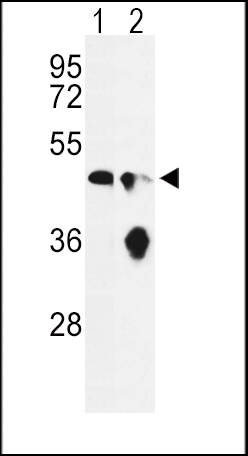
Western blot analysis of ENO1 Antibody (Center) in Ramos(lane 1), HL-60(lane 2) cell line lysates (35ug/lane). ENO1 (arrow) was detected using the purified Pab.Application
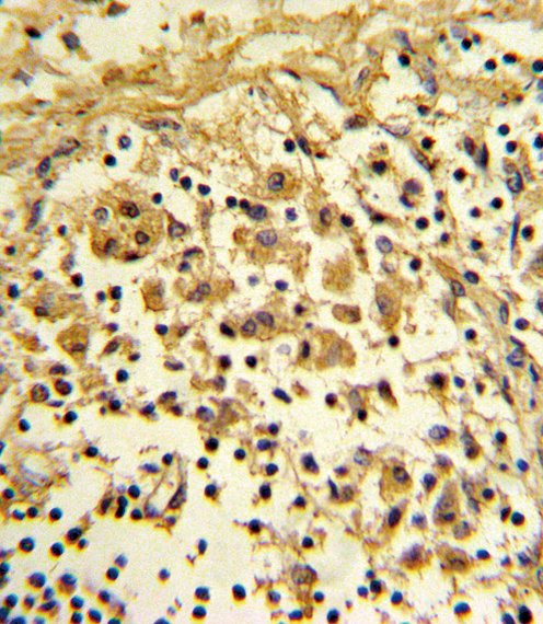
Formalin-fixed and paraffin-embedded human lymph reacted with ENO1 Antibody (Center), which was peroxidase-conjugated to the secondary antibody, followed by DAB staining. This data demonstrates the use of this antibody for immunohistochemistry; clinical relevance has not been evaluated.Application
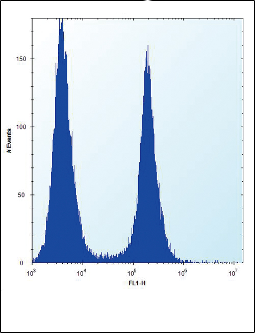
ENO1 Antibody (Center) flow cytometric analysis of Hela cells (right histogram) compared to a negative control cell (left histogram).FITC-conjugated donkey-anti-rabbit secondary antibodies were used for the analysis.Application
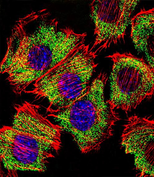
Fluorescent confocal image of C2C12 cell stained with ENO1 Antibody (Center)(Cat#AP6526c).C2C12 cells were fixed with 4% PFA (20 min), permeabilized with Triton X-100 (0.1%, 10 min), then incubated with ENO1 primary antibody (1:25, 1 h at 37℃). For secondary antibody, Alexa Fluor® 488 conjugated donkey anti-rabbit antibody (green) was used (1:400, 50 min at 37℃).Cytoplasmic actin was counterstained with Alexa Fluor® 555 (red) conjugated Phalloidin (7units/ml, 1 h at 37℃). Nuclei were counterstained with DAPI (blue) (10 µg/ml, 10 min). ENO1 immunoreactivity is localized to Cytoplasm significantly.| Product Name | ENO1 Antibody (Center) |
|---|---|
| Antibody Type | Primary Antibodies |
| Antigen Alias | ENO1; ENO1L1; MBPB1; MPB1; Alpha-enolase; 2-phospho-D-glycerate hydro-lyase; C-myc promoter-binding protein; Enolase 1; MBP-1; MPB-1; Non-neural enolase; Phosphopyruvate hydratase; Plasminogen-binding protein |
| Clonality | Polyclonal |
|---|---|
| Isotype | Ig |
| Host Species | Rabbit |
| Tested Applications | WBIHCFCIF |
| WB:1:100~500 IHC |
|
| Species Reactivity | Human |
| Concentration | 1mg/ml |
| Gene Synonyms | ENO1L1 MBPB1 MPB1 |
|---|---|
| Alternative Names | ENO1 ENO1L1 MBPB1 MPB1 Alpha-enolase 2-phospho-D-glycerate hydro-lyase C-myc promoter-binding protein Enolase 1 MBP-1 MPB-1 Non-neural enolase Phosphopyruvate hydratase Plasminogen-binding protein |
| Molecular Weight(MW) | 47169 Da |
| Function | Multifunctional enzyme that, as well as its role in glycolysis, plays a part in various processes such as growth control, hypoxia tolerance and allergic responses. May also function in the intravascular and pericellular fibrinolytic system due to its ability to serve as a receptor and activator of plasminogen on the cell surface of several cell-types such as leukocytes and neurons. Stimulates immunoglobulin production |
| Tissue Specificity | The alpha/alpha homodimer is expressed in embryo and in most adult tissues. The alpha/beta heterodimer and the beta/beta homodimer are found in striated muscle, and the alpha/gamma heterodimer and the gamma/gamma homodimer in neurons |
| Cellular Localization | Cytoplasm. Cell membrane. Cytoplasm, myofibril, sarcomere, M line. Note=Can translocate to the plasma membrane in either the homodimeric (alpha/alpha) or heterodimeric (alpha/gamma) form. ENO1 is localized to the M line |
| Entrez Gene | 2023 |
|---|
Application
Western blot analysis of ENO1 Antibody (Center) in Ramos(lane 1), HL-60(lane 2) cell line lysates (35ug/lane). ENO1 (arrow) was detected using the purified Pab.
Application
Formalin-fixed and paraffin-embedded human lymph reacted with ENO1 Antibody (Center), which was peroxidase-conjugated to the secondary antibody, followed by DAB staining. This data demonstrates the use of this antibody for immunohistochemistry; clinical relevance has not been evaluated.
Application
ENO1 Antibody (Center) flow cytometric analysis of Hela cells (right histogram) compared to a negative control cell (left histogram).FITC-conjugated donkey-anti-rabbit secondary antibodies were used for the analysis.
Application
Fluorescent confocal image of C2C12 cell stained with ENO1 Antibody (Center)(Cat#AP6526c).C2C12 cells were fixed with 4% PFA (20 min), permeabilized with Triton X-100 (0.1%, 10 min), then incubated with ENO1 primary antibody (1:25, 1 h at 37℃). For secondary antibody, Alexa Fluor® 488 conjugated donkey anti-rabbit antibody (green) was used (1:400, 50 min at 37℃).Cytoplasmic actin was counterstained with Alexa Fluor® 555 (red) conjugated Phalloidin (7units/ml, 1 h at 37℃). Nuclei were counterstained with DAPI (blue) (10 µg/ml, 10 min). ENO1 immunoreactivity is localized to Cytoplasm significantly.| Application Notes | WB:1:100~500 IHC |
|---|
| Form | Liquid |
|---|---|
| Storage Instructions | For short-term storage, store at 4° C. For long-term storage, aliquot and store at -20ºC or below. Avoid multiple freeze-thaw cycles. |
| Storage Buffer | Purified polyclonal antibody supplied in PBS with 0.09% (W/V) sodium azide. This antibody is prepared by Saturated Ammonium Sulfate (SAS) precipitation followed by dialysis against PBS. |


