Application
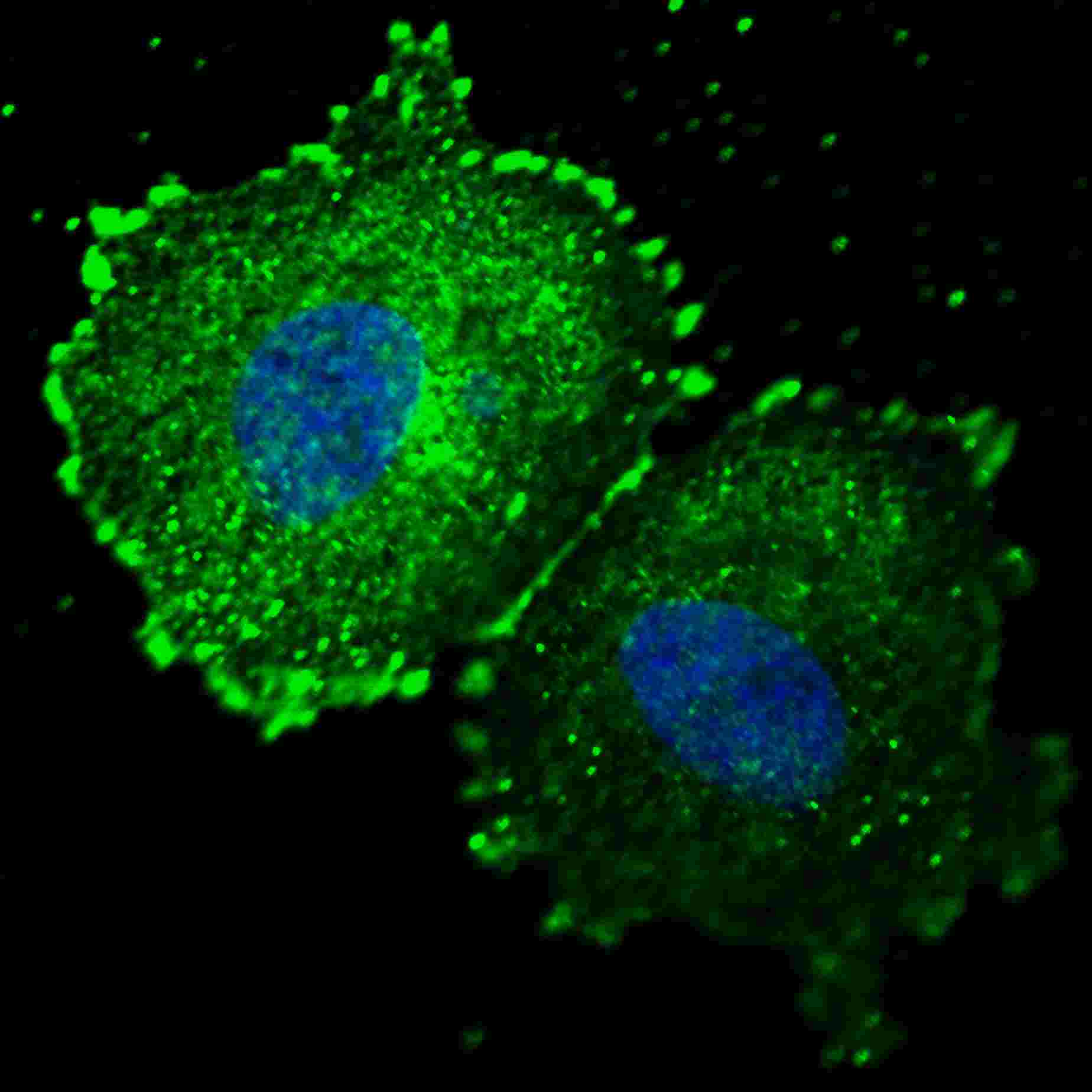
Fluorescent confocal image of MCF7 cells stained with phospho-ERBB2-Y1005 antibody. MCF7 cells were fixed with 4% PFA (20 min), permeabilized with Triton X-100 (0.2%, 30 min). Cells were then incubated with AP3781s phospho-ERBB2- Y1005 primary antibody (1:100, 2 h at room temperature). For secondary antibody, Alexa Fluor® 488 conjugated donkey anti-rabbit antibody (green) was used (1:1000, 1h). Nuclei were counterstained with Hoechst 33342 (blue) (10 μg/ml, 5 min). Note the highly specific localization of the phospho-ERBB2-Y1005 to the plasma membrane and cytoplasm.Application
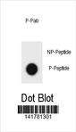
Dot blot analysis of ERBB2 Antibody (Phospho Y1005) Phospho-specific Pab on nitrocellulose membrane. 50ng of Phospho-peptide or Non Phospho-peptide per dot were adsorbed. Antibody working concentrations are 0.6ug per ml.| Product Name | ERBB2 Antibody (Phospho Y1005) |
|---|---|
| Antibody Type | Primary Antibodies |
| Modification | p-Y1005 |
| Clonality | Polyclonal |
|---|---|
| Isotype | Ig |
| Host Species | Rabbit |
| Tested Applications | IF,DB |
| IF:1:100 DB |
|
| Species Reactivity | Human |
| Concentration | 1mg/ml |
| Gene Synonyms | HER2, MLN19, NEU, NGL |
|---|---|
| Alternative Names | ERBB2 HER2 MLN19 NEU NGL Receptor tyrosine-protein kinase erbB-2 Metastatic lymph node gene 19 protein Proto-oncogene Neu Proto-oncogene c-ErbB-2 Tyrosine kinase-type cell surface receptor HER2 p185erbB2 CD_antigen=CD340 Flags: Precursor |
| Molecular Weight(MW) | 137910 Da |
| Function | Protein tyrosine kinase that is part of several cell surface receptor complexes, but that apparently needs a coreceptor for ligand binding. Essential component of a neuregulin-receptor complex, although neuregulins do not interact with it alone. GP30 is a potential ligand for this receptor. Regulates outgrowth and stabilization of peripheral microtubules (MTs). Upon ERBB2 activation, the MEMO1-RHOA-DIAPH1 signaling pathway elicits the phosphorylation and thus the inhibition of GSK3B at cell membrane This prevents the phosphorylation of APC and CLASP2, allowing its association with the cell membrane. In turn, membrane-bound APC allows the localization of MACF1 to the cell membrane, which is required for microtubule capture and stabilization |
| Tissue Specificity | Expressed in a variety of tumor tissues including primary breast tumors and tumors from small bowel, esophagus, kidney and mouth |
| Cellular Localization | Isoform 1: Cell membrane; Single-pass type I membrane protein. Cytoplasm, perinuclear region. Nucleus Note=Translocation to the nucleus requires endocytosis, probably endosomal sorting and is mediated by importin beta-1/KPNB1 Isoform 3: Cytoplasm. Nucleus. |
| Entrez Gene | 2064 |
|---|---|
| Nucleotide Accession | NP_001005862.1 |
Application
Fluorescent confocal image of MCF7 cells stained with phospho-ERBB2-Y1005 antibody. MCF7 cells were fixed with 4% PFA (20 min), permeabilized with Triton X-100 (0.2%, 30 min). Cells were then incubated with AP3781s phospho-ERBB2- Y1005 primary antibody (1:100, 2 h at room temperature). For secondary antibody, Alexa Fluor® 488 conjugated donkey anti-rabbit antibody (green) was used (1:1000, 1h). Nuclei were counterstained with Hoechst 33342 (blue) (10 μg/ml, 5 min). Note the highly specific localization of the phospho-ERBB2-Y1005 to the plasma membrane and cytoplasm.
Application
Dot blot analysis of ERBB2 Antibody (Phospho Y1005) Phospho-specific Pab on nitrocellulose membrane. 50ng of Phospho-peptide or Non Phospho-peptide per dot were adsorbed. Antibody working concentrations are 0.6ug per ml.| Application Notes | IF:1:100 DB |
|---|
| Form | Liquid |
|---|---|
| Storage Instructions | For short-term storage, store at 4° C. For long-term storage, aliquot and store at -20ºC or below. Avoid multiple freeze-thaw cycles. |
| Storage Buffer | Purified polyclonal antibody supplied in PBS with 0.09% (W/V) sodium azide. This antibody is purified through a protein A column, followed by two-step phosphospecific peptide affinity purification. |


