Application
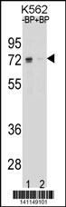
Western blot analysis of ESR1 isoform1 Antibody (C-term) Pab pre-incubated without(lane 1) and with(lane 2) blocking peptide in K562 cell line lysate. ESR1 isoform1 Antibody (C-term)(arrow) was detected using the purified Pab (1:60/250 dilution).Application
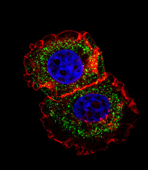
Fluorescent confocal image of MCF-7 cell stained with ESR1 isoform1 Antibody (C-term)(Cat#AP19616b). MCF-7 cells were fixed with 4% PFA (20 min), permeabilized with Triton X-100 (0.1%, 10 min), then incubated with ESR1 isoform1 primary antibody (1:25, 1 h at 37℃). For secondary antibody, Alexa Fluor® 488 conjugated donkey anti-rabbit antibody (green) was used (1:400, 50 min at 37℃).Cytoplasmic actin was counterstained with Alexa Fluor® 555 (red) conjugated Phalloidin (7units/ml, 1 h at 37℃). Nuclei were counterstained with DAPI (blue) (10 µg/ml, 10 min).ESR1 isoform1 immunoreactivity is localized to cytoplasm significantly.| Product Name | ESR1 isoform1 (C-term) |
|---|---|
| Antibody Type | Primary Antibodies |
| Clonality | Polyclonal |
|---|---|
| Isotype | Ig |
| Host Species | Rabbit |
| Tested Applications | WB,IF |
| WB:1:100~500 IF |
|
| Species Reactivity | Human |
| Concentration | 1mg/ml |
| Gene Synonyms | ESR, NR3A1 |
|---|---|
| Alternative Names | ESR1 ESR NR3A1 Estrogen receptor ER-alpha Estradiol receptor Nuclear receptor subfamily 3 group A member 1 |
| Molecular Weight(MW) | 66216 Da |
| Function | Nuclear hormone receptor. The steroid hormones and their receptors are involved in the regulation of eukaryotic gene expression and affect cellular proliferation and differentiation in target tissues. Ligand-dependent nuclear transactivation involves either direct homodimer binding to a palindromic estrogen response element (ERE) sequence or association with other DNA- binding transcription factors, such as AP-1/c-Jun, c-Fos, ATF-2, Sp1 and Sp3, to mediate ERE-independent signaling. Ligand binding induces a conformational change allowing subsequent or combinatorial association with multiprotein coactivator complexes through LXXLL motifs of their respective components. Mutual transrepression occurs between the estrogen receptor (ER) and NF- kappa-B in a cell-type specific manner. Decreases NF-kappa-B DNA- binding activity and inhibits NF-kappa-B-mediated transcription from the IL6 promoter and displace RELA/p65 and associated coregulators from the promoter. Recruited to the NF-kappa-B response element of the CCL2 and IL8 promoters and can displace CREBBP. Present with NF-kappa-B components RELA/p65 and NFKB1/p50 on ERE sequences. Can also act synergistically with NF-kappa-B to activate transcription involving respective recruitment adjacent response elements; the function involves CREBBP. Can activate the transcriptional activity of TFF1. Also mediates membrane-initiated estrogen signaling involving various kinase cascades. Isoform 3 is involved in activation of NOS3 and endothelial nitric oxide production. Isoforms lacking one or several functional domains are thought to modulate transcriptional activity by competitive ligand or DNA binding and/or heterodimerization with the full length receptor. Isoform 3 can bind to ERE and inhibit isoform 1 |
| Tissue Specificity | Widely expressed. Isoform 3 is not expressed in the pituitary gland |
| Cellular Localization | Isoform 1: Nucleus. Cytoplasm. Cell membrane; Peripheral membrane protein; Cytoplasmic side. Note=A minor fraction is associated with the inner membrane Nucleus. Golgi apparatus. Cell membrane. Note=Colocalizes with ZDHHC7 and ZDHHC21 in the Golgi apparatus where most probably palmitoylation occurs. Associated with the plasma membrane when palmitoylated |
| Entrez Gene | 2099 |
|---|---|
| Nucleotide Accession | NP_000116.2 |
Application
Western blot analysis of ESR1 isoform1 Antibody (C-term) Pab pre-incubated without(lane 1) and with(lane 2) blocking peptide in K562 cell line lysate. ESR1 isoform1 Antibody (C-term)(arrow) was detected using the purified Pab (1:60/250 dilution).
Application
Fluorescent confocal image of MCF-7 cell stained with ESR1 isoform1 Antibody (C-term)(Cat#AP19616b). MCF-7 cells were fixed with 4% PFA (20 min), permeabilized with Triton X-100 (0.1%, 10 min), then incubated with ESR1 isoform1 primary antibody (1:25, 1 h at 37℃). For secondary antibody, Alexa Fluor® 488 conjugated donkey anti-rabbit antibody (green) was used (1:400, 50 min at 37℃).Cytoplasmic actin was counterstained with Alexa Fluor® 555 (red) conjugated Phalloidin (7units/ml, 1 h at 37℃). Nuclei were counterstained with DAPI (blue) (10 µg/ml, 10 min).ESR1 isoform1 immunoreactivity is localized to cytoplasm significantly.| Application Notes | WB:1:100~500 IF |
|---|
| Form | Liquid |
|---|---|
| Storage Instructions | For short-term storage, store at 4° C. For long-term storage, aliquot and store at -20ºC or below. Avoid multiple freeze-thaw cycles. |
| Storage Buffer | Purified polyclonal antibody supplied in PBS with 0.09% (W/V) sodium azide. This antibody is purified through a protein A column, followed by peptide affinity purification. |


