Application
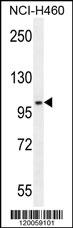
GABBR1 Antibody (Center) western blot analysis in NCI-H460 cell line lysates (35ug/lane).This demonstrates the GABBR1 antibody detected the GABBR1 protein (arrow).Application
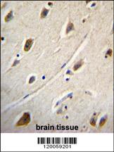
GABBR1 Antibody (Center) immunohistochemistry analysis in formalin fixed and paraffin embedded human brain tissue followed by peroxidase conjugation of the secondary antibody and DAB staining.This data demonstrates the use of GABBR1 Antibody (Center) for immunohistochemistry. Clinical relevance has not been evaluated.Application
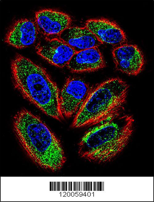
Confocal immunofluorescent analysis of GABBR1 Antibody (Center)(Cat#AP11234c) with NCI-H460 cell followed by Alexa Fluor 488-conjugated goat anti-rabbit lgG (green). Actin filaments have been labeled with Alexa Fluor 555 phalloidin (red).DAPI was used to stain the cell nuclear (blue).Application
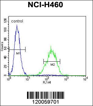
GABBR1 Antibody (Center) flow cytometric analysis of NCI-H460 cells (right histogram) compared to a negative control cell (left histogram).FITC-conjugated goat-anti-rabbit secondary antibodies were used for the analysis.| Product Name | GABBR1 Antibody (Center) |
|---|---|
| Antibody Type | Primary Antibodies |
| Clonality | Polyclonal |
|---|---|
| Isotype | Ig |
| Host Species | Rabbit |
| Tested Applications | WBIHCIFFC |
| WB:1:100~500 IHC |
|
| Species Reactivity | Human |
| Concentration | 1mg/ml |
| Gene Synonyms | GPRC3A |
|---|---|
| Alternative Names | GABBR1 GPRC3A Gamma-aminobutyric acid type B receptor subunit 1 |
| Molecular Weight(MW) | 108320 Da |
| Function | Receptor for GABA. The activity of this receptor is mediated by G-proteins that inhibit adenylyl cyclase activity, stimulates phospholipase A2, activates potassium channels, inactivates voltage-dependent calcium-channels and modulates inositol phospholipids hydrolysis. Plays a critical role in the fine-tuning of inhibitory synaptic transmission. Pre-synaptic GABA-B-R inhibit neurotransmitter release by down-regulating high- voltage activated calcium channels, whereas postsynaptic GABA-B-R decrease neuronal excitability by activating a prominent inwardly rectifying potassium (Kir) conductance that underlies the late inhibitory postsynaptic potentials. Not only implicated in synaptic inhibition but also in hippocampal long-term potentiation, slow wave sleep, muscle relaxation and antinociception. Activated by (-)-baclofen, cgp27492 and blocked by phaclofen |
| Tissue Specificity | Highly expressed in brain and weakly in heart, small intestine and uterus. Isoform 1A is mostly expressed in granular cell and molecular layer. Isoform 1B is mostly expressed in Purkinje cells. Isoform 1E is predominantly expressed in peripheral tissues as kidney, lung, trachea, colon, small intestine, stomach, bone marrow, thymus and mammary gland |
| Cellular Localization | Cell membrane; Multi-pass membrane protein. Cell junction, synapse, postsynaptic cell membrane; Multi-pass membrane protein. Note=Colocalizes with ATF4 in hippocampal neuron dendritic membranes (By similarity). Moreover coexpression of GABA-B-R1 and GABA-B-R2 appears to be a prerequisite for maturation and transport of GABA-B-R1 to the plasma membrane |
Application
GABBR1 Antibody (Center) western blot analysis in NCI-H460 cell line lysates (35ug/lane).This demonstrates the GABBR1 antibody detected the GABBR1 protein (arrow).
Application
GABBR1 Antibody (Center) immunohistochemistry analysis in formalin fixed and paraffin embedded human brain tissue followed by peroxidase conjugation of the secondary antibody and DAB staining.This data demonstrates the use of GABBR1 Antibody (Center) for immunohistochemistry. Clinical relevance has not been evaluated.
Application
Confocal immunofluorescent analysis of GABBR1 Antibody (Center)(Cat#AP11234c) with NCI-H460 cell followed by Alexa Fluor 488-conjugated goat anti-rabbit lgG (green). Actin filaments have been labeled with Alexa Fluor 555 phalloidin (red).DAPI was used to stain the cell nuclear (blue).
Application
GABBR1 Antibody (Center) flow cytometric analysis of NCI-H460 cells (right histogram) compared to a negative control cell (left histogram).FITC-conjugated goat-anti-rabbit secondary antibodies were used for the analysis.| Application Notes | WB:1:100~500 IHC |
|---|
| Form | Liquid |
|---|---|
| Storage Instructions | For short-term storage, store at 4° C. For long-term storage, aliquot and store at -20ºC or below. Avoid multiple freeze-thaw cycles. |
| Storage Buffer | Purified polyclonal antibody supplied in PBS with 0.09% (W/V) sodium azide. This antibody is purified through a protein A column, followed by peptide affinity purification. |


