Application
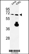
Western blot analysis of KLF4 Antibody in Jurkat, K562 cell line lysates (35ug/lane). KLF4 (arrow) was detected using the purified Pab.Application
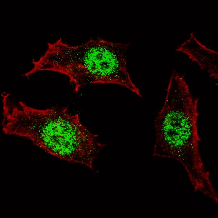
Fluorescent confocal image of HeLa cells stained with AP2725e KLF4 antibody. HeLa cells were fixed with 4% PFA (20 min), permeabilized with Triton X-100 (0.2%, 30 min), then incubated with AP2725e KLF4 primary antibody (1:100, 2 h at room temperature). For secondary antibody, Alexa Fluor® 488 conjugated donkey anti-rabbit antibody (green) was used (1:1000, 1h). Cytoplasmic actin was counterstained with Alexa Fluor® 555 (red) conjugated Phalloidin (5.25 μM, 25 min). KLF4 immunoreactivity is localized specifically to the nuclei in HeLa cells.Application
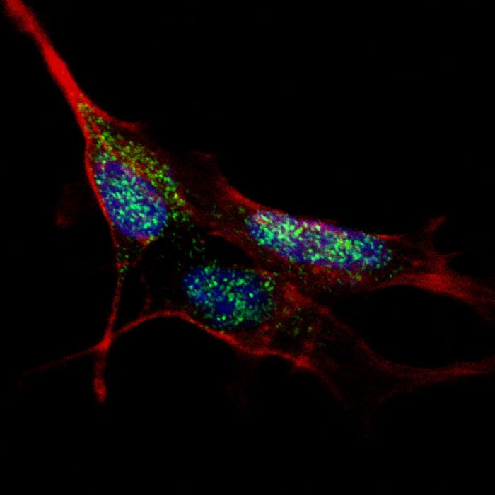
Fluorescent confocal image of SY5Y cells stained with AP2725e KLF4 antibody. SY5Y cells were fixed with 4% PFA (20 min), permeabilized with Triton X-100 (0.2%, 30 min), then incubated with AP2725e KLF4 primary antibody (1:100, 2 h at room temperature). For secondary antibody, Alexa Fluor® 488 conjugated donkey anti-rabbit antibody (green) was used (1:1000, 1h). Cytoplasmic actin was counterstained with Alexa Fluor® 555 (red) conjugated Phalloidin (5.25 μM, 25 min). Nuclei were counterstained with Hoechst 33342 (blue) (10 µg/ml, 3 min). KLF4 immunoreactivity is localized to the nuclei in SY5Y cells.Application
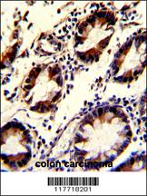
Formalin-fixed and paraffin-embedded human colon carcinoma with KLF4 Antibody, which was peroxidase-conjugated to the secondary antibody, followed by DAB staining. This data demonstrates the use of this antibody for immunohistochemistry; clinical relevance has not been evaluated.| Product Name | KLF4 Antibody |
|---|---|
| Antibody Type | Primary Antibodies |
| Antigen Alias | KLF4; EZF; GKLF; Krueppel-like factor 4; Epithelial zinc finger protein EZF; Gut-enriched krueppel-like factor |
| Clonality | Polyclonal |
|---|---|
| Isotype | Ig |
| Host Species | Rabbit |
| Tested Applications | WBIFIHC |
| WB:1:50~100 IF |
|
| Species Reactivity | Human |
| Concentration | 1mg/ml |
| Gene Synonyms | EZF GKLF |
|---|---|
| Alternative Names | KLF4 EZF GKLF Krueppel-like factor 4 Epithelial zinc finger protein EZF Gut-enriched krueppel-like factor |
| Molecular Weight(MW) | 54671 Da |
| Function | Transcription factor; can act both as activator and as repressor. Binds the 5'-CACCC-3' core sequence. Binds to the promoter region of its own gene and can activate its own transcription. Regulates the expression of key transcription factors during embryonic development. Plays an important role in maintaining embryonic stem cells, and in preventing their differentiation. Required for establishing the barrier function of the skin and for postnatal maturation and maintenance of the ocular surface. Involved in the differentiation of epithelial cells and may also function in skeletal and kidney development Contributes to the down-regulation of p53/TP53 transcription |
| Tissue Specificity | This KLF4 antibody is generated from rabbits immunized with a his tag recombinant protein of human KLF4. |
| Cellular Localization | Nucleus. |
Application
Western blot analysis of KLF4 Antibody in Jurkat, K562 cell line lysates (35ug/lane). KLF4 (arrow) was detected using the purified Pab.
Application
Fluorescent confocal image of HeLa cells stained with AP2725e KLF4 antibody. HeLa cells were fixed with 4% PFA (20 min), permeabilized with Triton X-100 (0.2%, 30 min), then incubated with AP2725e KLF4 primary antibody (1:100, 2 h at room temperature). For secondary antibody, Alexa Fluor® 488 conjugated donkey anti-rabbit antibody (green) was used (1:1000, 1h). Cytoplasmic actin was counterstained with Alexa Fluor® 555 (red) conjugated Phalloidin (5.25 μM, 25 min). KLF4 immunoreactivity is localized specifically to the nuclei in HeLa cells.
Application
Fluorescent confocal image of SY5Y cells stained with AP2725e KLF4 antibody. SY5Y cells were fixed with 4% PFA (20 min), permeabilized with Triton X-100 (0.2%, 30 min), then incubated with AP2725e KLF4 primary antibody (1:100, 2 h at room temperature). For secondary antibody, Alexa Fluor® 488 conjugated donkey anti-rabbit antibody (green) was used (1:1000, 1h). Cytoplasmic actin was counterstained with Alexa Fluor® 555 (red) conjugated Phalloidin (5.25 μM, 25 min). Nuclei were counterstained with Hoechst 33342 (blue) (10 µg/ml, 3 min). KLF4 immunoreactivity is localized to the nuclei in SY5Y cells.
Application
Formalin-fixed and paraffin-embedded human colon carcinoma with KLF4 Antibody, which was peroxidase-conjugated to the secondary antibody, followed by DAB staining. This data demonstrates the use of this antibody for immunohistochemistry; clinical relevance has not been evaluated.| Application Notes | WB:1:50~100 IF |
|---|
| Form | Liquid |
|---|---|
| Storage Instructions | For short-term storage, store at 4° C. For long-term storage, aliquot and store at -20ºC or below. Avoid multiple freeze-thaw cycles. |
| Storage Buffer | Purified polyclonal antibody supplied in PBS with 0.09% (W/V) sodium azide. This antibody is prepared by Saturated Ammonium Sulfate (SAS) precipitation followed by dialysis against PBS. |


