Application
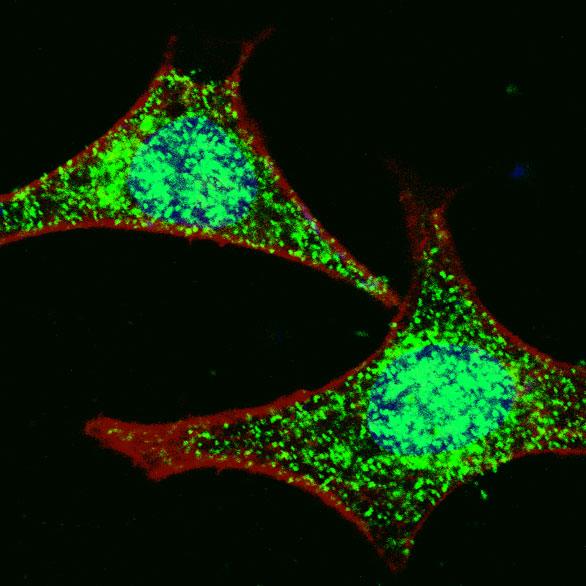
Fluorescent confocal image of HeLa cells stained with LGR5 (loop2) antibody. HeLa cells were fixed with 4% PFA (20 min), permeabilized with Triton X-100 (0.2%, 30 min). Cells were then incubated with AP2745d LGR5 (loop2) primary antibody (1:100, 2 h at room temperature). For secondary antibody, Alexa Fluor® 488 conjugated donkey anti-rabbit antibody (green) was used (1:1000, 1h). Nuclei were counterstained with Hoechst 33342 (blue) (10 μg/ml, 5 min). Note the highly specific localization of the LGR5 immunosignal to the nucleus and cytoplasm, supported by Human Protein Atlas Data (http://www.proteinatlas.org/ENSG00000139292).Application
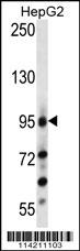
LGR5/GPR49 Antibody (loop2) western blot analysis in HepG2 cell line lysates (35ug/lane).This demonstrates the LGR5/GPR49 antibody detected the LGR5/GPR49 protein (arrow).Application
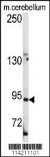
Western blot analysis of anti-LGR5/GPR49 Antibody (loop2) in mouse cerebellum tissue lysates (35ug/lane).LGR5/GPR49(arrow) was detected using the purified Pab.Application
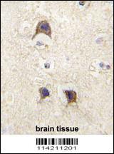
Formalin-fixed and paraffin-embedded human brain tissue reacted with LGR5/GPR49 antibody (loop2) , which was peroxidase-conjugated to the secondary antibody, followed by DAB staining. This data demonstrates the use of this antibody for immunohistochemistry; clinical relevance has not been evaluated.Application
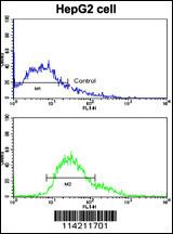
Flow cytometric analysis of HepG2 cells using LGR5/GPR49 Antibody (loop2) (bottom histogram) compared to a negative control cell (top histogram). FITC-conjugated goat-anti-rabbit secondary antibodies were used for the analysis.| Product Name | LGR5 Antibody (loop2) |
|---|---|
| Antibody Type | Primary Antibodies |
| Antigen Alias | LGR5; GPR49; GPR67; Leucine-rich repeat-containing G-protein coupled receptor 5; G-protein coupled receptor 49; G-protein coupled receptor 67; G-protein coupled receptor HG38 |
| Clonality | Polyclonal |
|---|---|
| Isotype | Ig |
| Host Species | Rabbit |
| Tested Applications | IFWBIHCFC |
| IF:1:100 WB |
|
| Species Reactivity | Human |
| Concentration | 1mg/ml |
| Gene Synonyms | GPR49 GPR67 |
|---|---|
| Alternative Names | LGR5 GPR49 GPR67 Leucine-rich repeat-containing G-protein coupled receptor 5 G-protein coupled receptor 49 G-protein coupled receptor 67 G-protein coupled receptor HG38 |
| Molecular Weight(MW) | 99998 Da |
| Function | Orphan receptor. Stem cell marker of the intestinal epithelium and the hair follicle. Target gene of Wnt signaling |
| Tissue Specificity | Expressed in skeletal muscle, placenta, spinal cord, and various region of brain. Expressed at the base of crypts in colonic and small mucosa stem cells. In premalignant cancer expression is not restricted to the cript base. Overexpressed in cancers of the ovary, colon and liver |
| Cellular Localization | Cell membrane; Multi-pass membrane protein. |
| Entrez Gene | 8549 |
|---|
Application
Fluorescent confocal image of HeLa cells stained with LGR5 (loop2) antibody. HeLa cells were fixed with 4% PFA (20 min), permeabilized with Triton X-100 (0.2%, 30 min). Cells were then incubated with AP2745d LGR5 (loop2) primary antibody (1:100, 2 h at room temperature). For secondary antibody, Alexa Fluor® 488 conjugated donkey anti-rabbit antibody (green) was used (1:1000, 1h). Nuclei were counterstained with Hoechst 33342 (blue) (10 μg/ml, 5 min). Note the highly specific localization of the LGR5 immunosignal to the nucleus and cytoplasm, supported by Human Protein Atlas Data (http://www.proteinatlas.org/ENSG00000139292).
Application
LGR5/GPR49 Antibody (loop2) western blot analysis in HepG2 cell line lysates (35ug/lane).This demonstrates the LGR5/GPR49 antibody detected the LGR5/GPR49 protein (arrow).
Application
Western blot analysis of anti-LGR5/GPR49 Antibody (loop2) in mouse cerebellum tissue lysates (35ug/lane).LGR5/GPR49(arrow) was detected using the purified Pab.
Application
Formalin-fixed and paraffin-embedded human brain tissue reacted with LGR5/GPR49 antibody (loop2) , which was peroxidase-conjugated to the secondary antibody, followed by DAB staining. This data demonstrates the use of this antibody for immunohistochemistry; clinical relevance has not been evaluated.
Application
Flow cytometric analysis of HepG2 cells using LGR5/GPR49 Antibody (loop2) (bottom histogram) compared to a negative control cell (top histogram). FITC-conjugated goat-anti-rabbit secondary antibodies were used for the analysis.| Application Notes | IF:1:100 WB |
|---|
| Form | Liquid |
|---|---|
| Storage Instructions | For short-term storage, store at 4° C. For long-term storage, aliquot and store at -20ºC or below. Avoid multiple freeze-thaw cycles. |
| Storage Buffer | Purified polyclonal antibody supplied in PBS with 0.09% (W/V) sodium azide. This antibody is prepared by Saturated Ammonium Sulfate (SAS) precipitation followed by dialysis against PBS. |


