Application
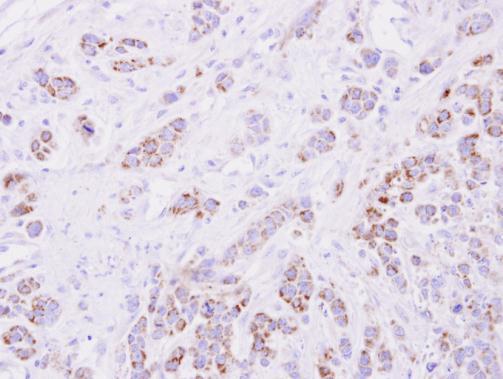
Immunohistochemical analysis of paraffin-embedded MDA-MB-468 xenograft, using 15-PGDH antibody at 1:500 dilution.Application
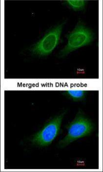
Immunofluorescence analysis of paraformaldehyde-fixed HeLa, using 15-PGDH antibody at 1:200 dilution.Application
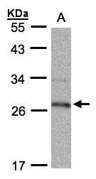
Sample (30 µg whole cell lysate) A:Raji 12% SDS PAGE diluted at 1:1000 The HRP-conjugated anti-rabbit IgG antibody was used to detect the primary antibody.IHC
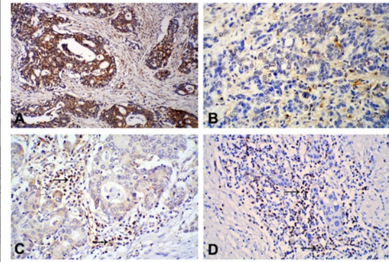
Positive staining of 15-PGDH protein was mainly found in the cytoplasm of gastric cancer (GC) tumor cells: (A) High/moderately differentiated GC, positive staining; (B) Poorly differentiated GC, negative staining. IHC staining of FOXP3 expression was mainly observed in the cytoplasm of lymphocytes in gastric cancer tissues: FOXP3 expression in high/moderately differentiated GC was lower (C) than in poorly differentiated GC (D) tissues. (×200).| Product Name | 15-PGDH antibody , C-term |
|---|---|
| Antibody Type | Primary Antibodies |
| Product description | Rabbit Polyclonal antibody to 15-PGDH (hydroxyprostaglandin dehydrogenase 15-(NAD))This gene encodes a member of the short-chain nonmetalloenzyme alcohol dehydrogenase protein family. The encoded enzyme is responsible for the metabolism of prostaglandins, which function in a variety of physiologic and cellular processes such as inflammation. Mutations in this gene result in primary autosomal recessive hypertrophic osteoarthropathy and cranioosteoarthropathy. Multiple transcript variants encoding different isoforms have been found for this gene. [provided by RefSeq] |
| Immunogen | Carrier-protein conjugated synthetic peptide encompassing a sequence within the C-terminus region of human 15-PGDH. The exact sequence is proprietary. |
| Clonality | Polyclonal |
|---|---|
| Isotype | IgG |
| Host Species | Rabbit |
| Tested Applications | ICC/IFIHC-PWB |
| *Optimal dilutions/concentrations should be determined by the researcher.
: |
|
| Species Reactivity | Human |
| Predicted Reactivity | PigPig (83%) |
| Concentration | 1mg/ |
| Purification | Affinity purified |
| Gene Synonyms | 15-PGDH antibody PGDH antibody PGDH1 antibody SDR36C1 antibody HPGD antibody prostaglandin dehydrogenase 1 antibody NAD+-dependent 15-hydroxyprostaglandin dehydrogenase antibody 15-hydroxyprostaglandin dehydrogenase [NAD+] antibody "short chain deh |
|---|---|
| Molecular Weight(MW) | 29 kDa (note) (4) |
Application
Immunohistochemical analysis of paraffin-embedded MDA-MB-468 xenograft, using 15-PGDH antibody at 1:500 dilution.
Application
Immunofluorescence analysis of paraformaldehyde-fixed HeLa, using 15-PGDH antibody at 1:200 dilution.
Application
Sample (30 µg whole cell lysate) A:Raji 12% SDS PAGE diluted at 1:1000 The HRP-conjugated anti-rabbit IgG antibody was used to detect the primary antibody.
IHC
Positive staining of 15-PGDH protein was mainly found in the cytoplasm of gastric cancer (GC) tumor cells: (A) High/moderately differentiated GC, positive staining; (B) Poorly differentiated GC, negative staining. IHC staining of FOXP3 expression was mainly observed in the cytoplasm of lymphocytes in gastric cancer tissues: FOXP3 expression in high/moderately differentiated GC was lower (C) than in poorly differentiated GC (D) tissues. (×200).| Positive Control | 293T , A431 , HeLaS3 , HepG2 , Molt-4 , Raji |
|---|---|
| Application Notes | *Optimal dilutions/concentrations should be determined by the researcher.
: |
| Form | Liquid |
|---|---|
| Storage Instructions | Keep as concentrated solution. Aliquot and store at -20ºC or below. Avoid multiple freeze-thaw cycles. |
| Storage Buffer | 1XPBS, 40% Glycerol (pH7). 0.01% Thimerosal was added as a preservative. |


