Application
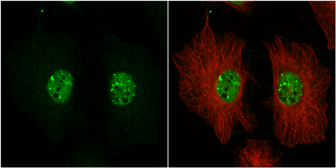
53BP1 antibody detects 53BP1 protein at nucleus by immunofluorescent analysis. Sample: HeLa cells were fixed in 4% paraformaldehyde at RT for 15 min. Green: 53BP1 protein stained by 53BP1 antibody diluted at 1:1000. Red: alpha Tubulin, a cytoskeleton marker, stained by alpha Tubulin antibody [GT114] diluted at 1:1000.Application
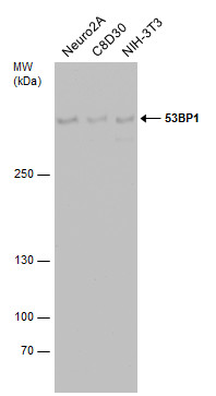
Various whole cell extracts (30 µg) were separated by 5% SDS-PAGE, and the membrane was blotted with 53BP1 antibody diluted at 1:500. The HRP-conjugated anti-rabbit IgG antibody was used to detect the primary antibody.Application
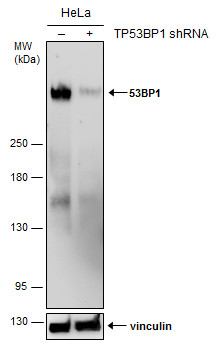
Non-transfected (–) and transfected (+) HeLa whole cell extracts (50 µg) were separated by 5% SDS-PAGE, and the membrane was blotted with 53BP1 antibody diluted at 1:500. The HRP-conjugated anti-rabbit IgG antibody was used to detect the primary antibody.Application
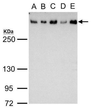
53BP1 antibody detects 53BP1 protein by western blot analysis. A. 30 µg 293T whole cell lysate/extract B. 30 µg A431 whole cell lysate/extract C. 30 µg HeLa whole cell lysate/extract D. 30 µg HepG2 whole cell lysate/extract E. 30 µg A375 whole cell lysate/extract 5% SDS-PAGE 53BP1 antibody dilution: 1:2000 The HRP-conjugated anti-rabbit IgG antibody was used to detect the primary antibody.| Product Name | 53BP1 antibody |
|---|---|
| Antibody Type | Primary Antibodies |
| Product description | Rabbit polyclonal antibody to human 53BP1 ( carboxy terminus )P53-binding protein 1 (53BP1) plays a critical role in tumor suppression and is a putative substrate of ATM kinase. Upon DNA damage, it is phosphorylated and relocalizes to the presumptive sites of damage, specifically, double-strand breaks. This also suggests a role in DNA repair, maintaining genomic stability. |
| Immunogen | Synthetic peptide corresponding to the carboxy-terminus of h53BP1, conjugated to a carrier protein. |
| Clonality | Polyclonal |
|---|---|
| Isotype | IgG |
| Host Species | Rabbit |
| Tested Applications | ICC/IFIHCWB |
| Suggested starting dilutions are as follows: WB: 1:500-1:3000, ICC/IF: 1:100-1:1000. Not yet tested in other applications. Optimal working dilutions should be determined experimentally by the end user.: |
|
| Species Reactivity | HumanMouse |
| Concentration | 1mg/ml (Please refer to the vial label for the specific concentration) |
| Purification | Affinity purified |
| Gene Synonyms | 53BP1 MGC138366 TP53BP1 p202 7158 605230 Q12888 FLJ41424 |
|---|---|
| Target | 53BP1 |
Application
53BP1 antibody detects 53BP1 protein at nucleus by immunofluorescent analysis. Sample: HeLa cells were fixed in 4% paraformaldehyde at RT for 15 min. Green: 53BP1 protein stained by 53BP1 antibody diluted at 1:1000. Red: alpha Tubulin, a cytoskeleton marker, stained by alpha Tubulin antibody [GT114] diluted at 1:1000.
Application
Various whole cell extracts (30 µg) were separated by 5% SDS-PAGE, and the membrane was blotted with 53BP1 antibody diluted at 1:500. The HRP-conjugated anti-rabbit IgG antibody was used to detect the primary antibody.
Application
Non-transfected (–) and transfected (+) HeLa whole cell extracts (50 µg) were separated by 5% SDS-PAGE, and the membrane was blotted with 53BP1 antibody diluted at 1:500. The HRP-conjugated anti-rabbit IgG antibody was used to detect the primary antibody.
Application
53BP1 antibody detects 53BP1 protein by western blot analysis. A. 30 µg 293T whole cell lysate/extract B. 30 µg A431 whole cell lysate/extract C. 30 µg HeLa whole cell lysate/extract D. 30 µg HepG2 whole cell lysate/extract E. 30 µg A375 whole cell lysate/extract 5% SDS-PAGE 53BP1 antibody dilution: 1:2000 The HRP-conjugated anti-rabbit IgG antibody was used to detect the primary antibody.| Positive Control | 293T , A431 , HeLa , HepG2 , A375 , Neuro 2A , C8D30 , NIH-3T3 , *HaCaT |
|---|---|
| Application Notes | Suggested starting dilutions are as follows: WB: 1:500-1:3000, ICC/IF: 1:100-1:1000. Not yet tested in other applications. Optimal working dilutions should be determined experimentally by the end user.: |
| Form | Liquid |
|---|---|
| Storage Instructions | Keep as concentrated solution. Aliquot and store at -20ºC or below. Avoid multiple freeze-thaw cycles. |
| Storage Buffer | PBS |


