Application
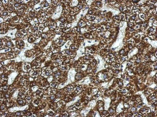
Immunohistochemical analysis of paraffin-embedded human hepatoma, using SBCAD antibody at 1:500 dilution.Application
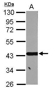
Sample (30 ug of whole cell lysate) A: HepG2 10% SDS PAGE diluted at 1:10000Application
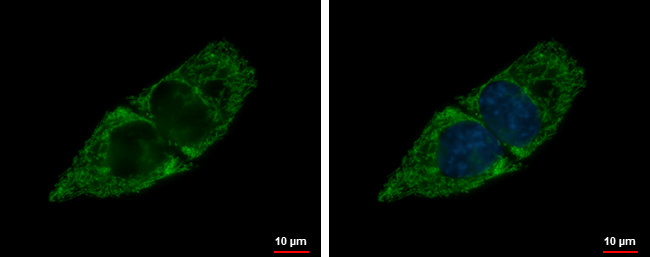
SBCAD antibody detects SBCAD protein at mitochondria by immunofluorescent analysis. Sample: HepG2 cells were fixed in 2% paraformaldehyde/culture medium at 37℃ for 30 min. Green: SBCAD protein stained by SBCAD antibody diluted at 1:5000. Blue: Hoechst 33342 staining.Application
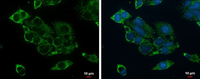
SBCAD antibody detects SBCAD protein at mitochondria by immunofluorescent analysis. Sample: Hep G2 cells were fixed in 2% paraformaldehyde/culture medium at 37oC for 30 min. Green: SBCAD protein stained by SBCAD antibody diluted at 1:500. Blue: Hoechst 33342 staining. Scale bar = 10 μm.| Product Name | ACADSB antibody |
|---|---|
| Antibody Type | Primary Antibodies |
| Product description | Rabbit Polyclonal antibody to ACADSB (acyl-CoA dehydrogenase, short/branched chain)Short/branched chain acyl-CoA dehydrogenase(ACADSB) is a member of the acyl-CoA dehydrogenase family of enzymes that catalyze the dehydrogenation of acyl-CoA derivatives in the metabolism of fatty acids or branch chained amino acids. Substrate specificity is the primary characteristic used to define members of this gene family. The ACADSB gene product has the greatest activity towards the short branched chain acyl-CoA derivative, (S)-2-methylbutyryl-CoA, but also reacts significantly with other 2-methyl branched chain substrates and with short straight chain acyl-CoAs. The cDNA encodes for a mitochondrial precursor protein which is cleaved upon mitochondrial import and predicted to yield a mature peptide of approximately 43.7-KDa. [provided by RefSeq] |
| Immunogen | Recombinant protein encompassing a sequence within the center region of human ACADSB. The exact sequence is proprietary. |
| Clonality | Polyclonal |
|---|---|
| Isotype | IgG |
| Host Species | Rabbit |
| Tested Applications | ICC/IFIHC-PWB |
| *Optimal dilutions/concentrations should be determined by the researcher.
: |
|
| Species Reactivity | Human |
| Predicted Reactivity | Rat, Rhesus MonkeyRat (81%), Rhesus Monkey (94%) |
| Concentration | 1mg/ml (Please refer to the vial label for the specific concentration) |
| Purification | Affinity purified |
| Gene Synonyms | 2-MEBCAD antibody ACAD7 antibody SBCAD antibody ACADSB antibody 2-methylbutyryl-coenzyme A dehydrogenase antibody "acyl-Coenzyme A dehydrogenase short/branched chain antibody" "short/branched chain specific acyl-CoA dehydrogenase mitochondrial anti |
|---|---|
| Molecular Weight(MW) | 47 kDa (note) (2) |
| Cellular Localization | Mitochondrion matrix |
Application
Immunohistochemical analysis of paraffin-embedded human hepatoma, using SBCAD antibody at 1:500 dilution.
Application
Sample (30 ug of whole cell lysate) A: HepG2 10% SDS PAGE diluted at 1:10000
Application
SBCAD antibody detects SBCAD protein at mitochondria by immunofluorescent analysis. Sample: HepG2 cells were fixed in 2% paraformaldehyde/culture medium at 37℃ for 30 min. Green: SBCAD protein stained by SBCAD antibody diluted at 1:5000. Blue: Hoechst 33342 staining.
Application
SBCAD antibody detects SBCAD protein at mitochondria by immunofluorescent analysis. Sample: Hep G2 cells were fixed in 2% paraformaldehyde/culture medium at 37oC for 30 min. Green: SBCAD protein stained by SBCAD antibody diluted at 1:500. Blue: Hoechst 33342 staining. Scale bar = 10 μm.| Positive Control | HepG2 |
|---|---|
| Application Notes | *Optimal dilutions/concentrations should be determined by the researcher.
: |
| Form | Liquid |
|---|---|
| Storage Instructions | Keep as concentrated solution. Aliquot and store at -20ºC or below. Avoid multiple freeze-thaw cycles. |
| Storage Buffer | 1XPBS, 20% Glycerol (pH7). 0.01% Thimerosal was added as a preservative. |


