Application
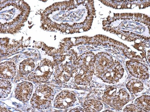
Aconitase 2 antibody [N1N3] detects Aconitase 2 protein at mitochondria on mouse intestine by immunohistochemical analysis. Sample: Paraffin-embedded mouse intestine. Aconitase 2 antibody [N1N3] dilution: 1:500.Application
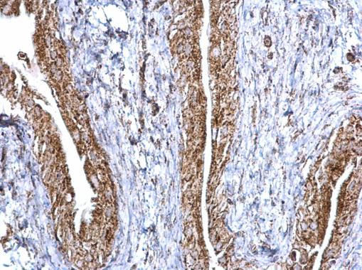
Aconitase 2 antibody [N1N3] detects Aconitase 2 protein at mitochondria on mouse urinary bladder by immunohistochemical analysis. Sample: Paraffin-embedded mouse urinary bladder. Aconitase 2 antibody [N1N3] dilution: 1:500.Application
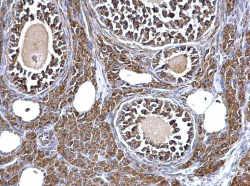
Aconitase 2 antibody [N1N3] detects Aconitase 2 protein at mitochondria on mouse kidney by immunohistochemical analysis. Sample: Paraffin-embedded mouse ovary. Aconitase 2 antibody [N1N3] dilution: 1:500.Application
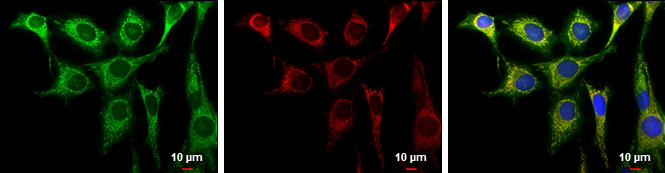
Aconitase 2 antibody [N1N3] detects Aconitase 2 protein at mitochondria by immunofluorescent analysis. Sample: HeLa cells were fixed in 2% paraformaldehyde/culture medium at 37oC for 30 min. Green: Aconitase 2 protein stained by Aconitase 2 antibody [N1N3] diluted at 1:1000. Red: MitoTrackerR Red CMXRos, a mitochondria tracker. Blue: Hoechst 33342 staining. Scale bar = 10 μm.Application
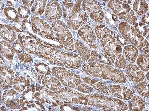
Aconitase 2 antibody [N1N3] detects Aconitase 2 protein at mitochondria on mouse liver by immunohistochemical analysis. Sample: Paraffin-embedded mouse kidney. Aconitase 2 antibody [N1N3] dilution: 1:500.Application
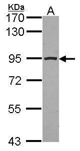
Sample (30 µg of whole cell lysate) A: JurKat 7.5% SDS PAGE Aconitase 2 antibody diluted at 1:1000 The HRP-conjugated anti-rabbit IgG antibody was used to detect the primary antibody.Application
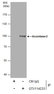
Immunoprecipitation of Aconitase 2 protein from HeLa whole cell extracts using 5 μg of Aconitase 2 antibody [N1N3] . Western blot analysis was performed using Aconitase 2 antibody [N1N3] . EasyBlot anti-Rabbit IgG was used as a secondary reagent.Application
Sample (20 µg of whole cell lysate) A: mouse brain 7.5% SDS PAGE diluted at 1:10000 The HRP-conjugated anti-rabbit IgG antibody was used to detect the primary antibody.Application
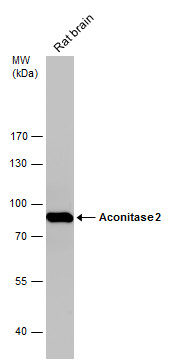
Rat tissue extract (50 µg) was separated by 7.5% SDS-PAGE, and the membrane was blotted with Aconitase 2 antibody [N1N3] diluted at 1:10000. The HRP-conjugated anti-rabbit IgG antibody was used to detect the primary antibody.Application
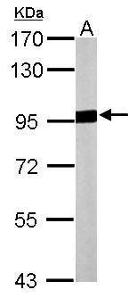
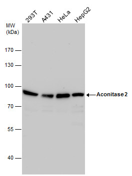
Aconitase 2 antibody detects Aconitase 2 protein by western blot analysis. Various whole cell extracts (30 µg) were separated by 7.5% SDS-PAGE, and the membrane was blotted with Aconitase 2 antibody diluted by 1:1000. The HRP-conjugated anti-rabbit IgG antibody was used to detect the primary antibody.| Product Name | Aconitase 2 antibody |
|---|---|
| Antibody Type | Primary Antibodies |
| Product description | Rabbit Polyclonal antibody to Aconitase 2 (aconitase 2, mitochondrial)The protein encoded by this gene belongs to the aconitase/IPM isomerase family. It is an enzyme that catalyzes the interconversion of citrate to isocitrate via cis-aconitate in the second step of the TCA cycle. This protein is encoded in the nucleus and functions in the mitochondrion. It was found to be one of the mitochondrial matrix proteins that are preferentially degraded by the serine protease 15(PRSS15), also known as Lon protease, after oxidative modification. [provided by RefSeq] |
| Immunogen | Recombinant protein encompassing a sequence within the center region of human Aconitase 2. The exact sequence is proprietary. |
| Clonality | Polyclonal |
|---|---|
| Isotype | IgG |
| Host Species | Rabbit |
| Tested Applications | ICC/IFIHC-PIPWB |
| *Optimal dilutions/concentrations should be determined by the researcher.
: |
|
| Species Reactivity | HumanMouseChickenRat |
| Predicted Reactivity | Zebrafish, Xenopus laevis, Pig, Rhesus Monkey, BovineZebrafish (91%), Xenopus laevis (94%), Pig (98%), Rhesus Monkey (98%), Bovine (97%) |
| Concentration | 1mg/ml (Please refer to the vial label for the specific concentration) |
| Purification | Affinity purified |
| Gene Synonyms | ACONM antibody MGC20605 antibody MGC33908 antibody ACO2 antibody citrate hydro-lyase antibody "aconitate hydratase mitochondrial antibody" "aconitase 2 mitochondrial antibody" |
|---|---|
| Molecular Weight(MW) | 85 kDa (note) (2) |
| Cellular Localization | Mitochondrion |
Application
Aconitase 2 antibody [N1N3] detects Aconitase 2 protein at mitochondria on mouse intestine by immunohistochemical analysis. Sample: Paraffin-embedded mouse intestine. Aconitase 2 antibody [N1N3] dilution: 1:500.
Application
Aconitase 2 antibody [N1N3] detects Aconitase 2 protein at mitochondria on mouse urinary bladder by immunohistochemical analysis. Sample: Paraffin-embedded mouse urinary bladder. Aconitase 2 antibody [N1N3] dilution: 1:500.
Application
Aconitase 2 antibody [N1N3] detects Aconitase 2 protein at mitochondria on mouse kidney by immunohistochemical analysis. Sample: Paraffin-embedded mouse ovary. Aconitase 2 antibody [N1N3] dilution: 1:500.
Application
Aconitase 2 antibody [N1N3] detects Aconitase 2 protein at mitochondria by immunofluorescent analysis. Sample: HeLa cells were fixed in 2% paraformaldehyde/culture medium at 37oC for 30 min. Green: Aconitase 2 protein stained by Aconitase 2 antibody [N1N3] diluted at 1:1000. Red: MitoTrackerR Red CMXRos, a mitochondria tracker. Blue: Hoechst 33342 staining. Scale bar = 10 μm.
Application
Aconitase 2 antibody [N1N3] detects Aconitase 2 protein at mitochondria on mouse liver by immunohistochemical analysis. Sample: Paraffin-embedded mouse kidney. Aconitase 2 antibody [N1N3] dilution: 1:500.
Application
Sample (30 µg of whole cell lysate) A: JurKat 7.5% SDS PAGE Aconitase 2 antibody diluted at 1:1000 The HRP-conjugated anti-rabbit IgG antibody was used to detect the primary antibody.
Application
Immunoprecipitation of Aconitase 2 protein from HeLa whole cell extracts using 5 μg of Aconitase 2 antibody [N1N3] . Western blot analysis was performed using Aconitase 2 antibody [N1N3] . EasyBlot anti-Rabbit IgG was used as a secondary reagent.
Application
Sample (20 µg of whole cell lysate) A: mouse brain 7.5% SDS PAGE diluted at 1:10000 The HRP-conjugated anti-rabbit IgG antibody was used to detect the primary antibody.
Application
Rat tissue extract (50 µg) was separated by 7.5% SDS-PAGE, and the membrane was blotted with Aconitase 2 antibody [N1N3] diluted at 1:10000. The HRP-conjugated anti-rabbit IgG antibody was used to detect the primary antibody.
Application
Aconitase 2 antibody detects Aconitase 2 protein by western blot analysis. Various whole cell extracts (30 µg) were separated by 7.5% SDS-PAGE, and the membrane was blotted with Aconitase 2 antibody diluted by 1:1000. The HRP-conjugated anti-rabbit IgG antibody was used to detect the primary antibody.| Positive Control | 293T , A431 , HeLa , HepG2 , JurKat , Mouse brain , rat brain |
|---|---|
| Application Notes | *Optimal dilutions/concentrations should be determined by the researcher.
: |
| Form | Liquid |
|---|---|
| Storage Instructions | Keep as concentrated solution. Aliquot and store at -20ºC or below. Avoid multiple freeze-thaw cycles. |
| Storage Buffer | 0.1M Tris, 0.1M Glycine, 20% Glycerol (pH7). 0.01% Thimerosal was added as a preservative. |


