Application
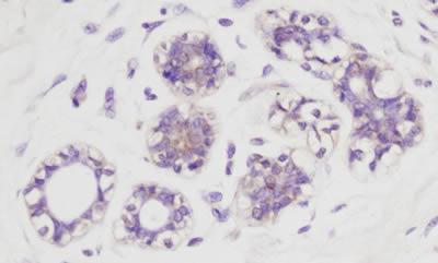
Rabbit anti-AKT pS473 was used at a 1:100 dilution to detect AKT by immunohistochemistry in normal human breast tissue. AKT is weakly phosphorylated in normal tissue in the breast. The phosphorylated AKT is clearly localized in the cytoplasm. Rabbit anti-AKT pS473 antibody was used with no pretreatment of tissue. Tissue was formalin-fixed and paraffin embedded.Application
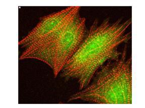
Immunofluorescence Confocal Microscopy of Rabbit anti-AKT pS473 antibody . Tissue: cardiomyocytes infected with adenovirus expressing with wild-type AKT. Fixation: 0.5% PFA. Antigen retrieval: not required. Primary antibody: AKT pS473 antibody at 1:100 for 1 h at RT. Secondary antibody: texas-red conjugated rabbit secondary antibody at 1:10,000 for 45 min at RT. Localization: AKT pS473 is nuclear. Staining: AKT pS473 as green fluorescent signal with texas-red conjugated phalloidin (red) to label filamentous actin.Application
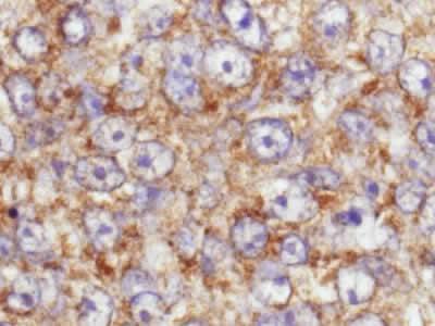
This image is a magnification of the accompanying breast tumor image.Application
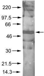
Rabbit anti-AKT pS473 was used at a 1:500 dilution to detect phosphorylated AKT by Western blot. A nuclear extract from cells infected with adenovirus expressing nuclear-targeted AKT kinase was used.Application
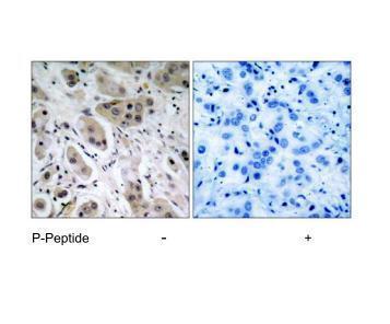
Immunohistochemical analysis of paraffin- embedded human breast carcinoma tissue, using Akt (phospho-Ser473) antibody .Application
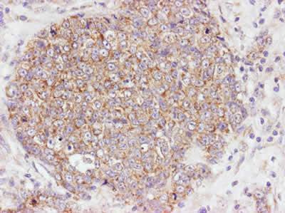
Rabbit anti-AKT pS473 was used at a 1:100 dilution to detect AKT by immunohistochemistry in human breast tumor tissue. The staining is much stronger than the weak basal level of phosphorylation in normal breast.Rabbit anti-AKT pS473 antibody was used with no pretreatment of tissue.Tissue was formalin-fixed and paraffin embedded.Application
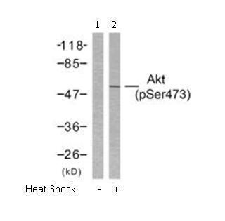
Western blot analysis of extract from HeLa cells untreated or treated with heat shock using Akt (phospho-Ser473) antibody ( Lane 1 and 2).| Product Name | AKT (phospho Ser473) antibody |
|---|---|
| Antibody Type | Primary Antibodies |
| Product description | Rabbit polyclonal to AKT (phospho S473)Akt (Protein Kinase B, PKB) is a key regulator of many signal transduction pathways (reviewed in ref 1). Its tight control over cell proliferation and cell viability are manifold; overexpression or inappropriate activation of Akt has been seen in many types of cancer. Akt mediates many of the downstream events of PI 3-kinase (a lipid kinase activated by growth factors, cytokines and insulin). PI 3-kinase recruits Akt to the membrane, where it is activated by PDK1 phosphorylation (reviewed in 2). Once phosphorylated, Akt dissociates from the membrane and phosphorylates targets in the cytoplasm and the cell nucleus. Akt has two main roles: (i) inhibition of apoptosis; (ii) promotion of proliferation. |
| Immunogen | Synthetic peptide: CHFPQFS p YSAS conjugated to KLH, corresponding to amino acids 460-480 of Human AKT. |
| Conjugation | Agarose |
| Modification | p-Ser473 |
| Clonality | Polyclonal |
|---|---|
| Isotype | IgG |
| Host Species | Rabbit |
| Tested Applications | ICC/IFIHC-PWB |
| For IHC-P: Use at a dilution of 1:50 - 1:200. No pre-treatment is required. For WB: Use at a dilution of 1:500 - 1:2000. Optimal dilutions/concentrations should be determined by the researcher.: |
|
| Species Reactivity | HumanMouseRat |
| Concentration | 1mg/ml (Please refer to the vial label for the specific concentration) |
| Purification | Affinity purified |
| Gene Synonyms | P31751 RAC 207 RAC-BETA MGC99656 208 PKBBETA PRKBB RACBETA 125853 164730 AKT2 AKT1 P31749 PRKBA RAC-ALPHA RACALPHA PKB AKT RAC BETA RAC ALPHA AKT (phospho S473) AKT (phospho Ser473) AKT (pS473) AKT (pSer473) AKT phospho S473 AKT p |
|---|---|
| Target | AKT |
| Molecular Weight(MW) | 53KDa (note) |
| Tissue Specificity | Akt (phospho-Ser473) antibody detects endogenous levels of Akt only when phosphorylated at serine 473. |
Application
Rabbit anti-AKT pS473 was used at a 1:100 dilution to detect AKT by immunohistochemistry in normal human breast tissue. AKT is weakly phosphorylated in normal tissue in the breast. The phosphorylated AKT is clearly localized in the cytoplasm. Rabbit anti-AKT pS473 antibody was used with no pretreatment of tissue. Tissue was formalin-fixed and paraffin embedded.
Application
Immunofluorescence Confocal Microscopy of Rabbit anti-AKT pS473 antibody . Tissue: cardiomyocytes infected with adenovirus expressing with wild-type AKT. Fixation: 0.5% PFA. Antigen retrieval: not required. Primary antibody: AKT pS473 antibody at 1:100 for 1 h at RT. Secondary antibody: texas-red conjugated rabbit secondary antibody at 1:10,000 for 45 min at RT. Localization: AKT pS473 is nuclear. Staining: AKT pS473 as green fluorescent signal with texas-red conjugated phalloidin (red) to label filamentous actin.
Application
This image is a magnification of the accompanying breast tumor image.
Application
Rabbit anti-AKT pS473 was used at a 1:500 dilution to detect phosphorylated AKT by Western blot. A nuclear extract from cells infected with adenovirus expressing nuclear-targeted AKT kinase was used.
Application
Immunohistochemical analysis of paraffin- embedded human breast carcinoma tissue, using Akt (phospho-Ser473) antibody .
Application
Rabbit anti-AKT pS473 was used at a 1:100 dilution to detect AKT by immunohistochemistry in human breast tumor tissue. The staining is much stronger than the weak basal level of phosphorylation in normal breast.Rabbit anti-AKT pS473 antibody was used with no pretreatment of tissue.Tissue was formalin-fixed and paraffin embedded.
Application
Western blot analysis of extract from HeLa cells untreated or treated with heat shock using Akt (phospho-Ser473) antibody ( Lane 1 and 2).| Application Notes | For IHC-P: Use at a dilution of 1:50 - 1:200. No pre-treatment is required. For WB: Use at a dilution of 1:500 - 1:2000. Optimal dilutions/concentrations should be determined by the researcher.: |
|---|
| Form | Liquid |
|---|---|
| Storage Instructions | Store vial at -20 °C or below prior to opening. Dilute only prior to immediate use. Aliquot contents and freeze at -20 °C or below. Avoid cycles of freezing and thawing. |
| Storage Buffer | Phosphate-buffered saline, pH 7.3, 50% Glycerol and 150mM NaCl containing 0.02% sodium azide |


