Application
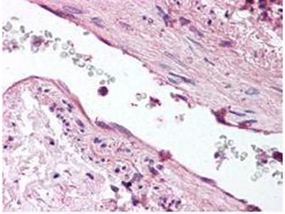
IHC-P of ANG1 antibody was diluted 1:500 to detect ANG1 in human lung tissue. The image shows the localization of antibody as the precipitated red signal, with a hematoxylin purple nuclear counter stain.Application
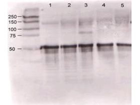
Rabbit anti-Ang-1 was used at a 1:500 dilution to detect mouse Ang-1 by western blot against supernatants of mouse angiopoietin-expressing endothelial cells. Lane 1 - wt endothelial cells. Lane 2 - mouse Ang-1 (clone 1-8) expressing cells. Lane 3 - mouse Ang-1 (clone 1-15) expressing cells. Lane 4 - mouse Ang-2 (clone 2-9) expressing cells. Approximately 20 µg of each lysate was used for 10% SDS-PAGE.| Product Name | Angiopoietin 1 antibody |
|---|---|
| Antibody Type | Primary Antibodies |
| Product description | Rabbit Polyclonal antibody to Angiopoietin 1Angiopoeitin-1 (Ang-1) and Antiopoietin-2 (Ang2) are important for development of the endothelium, by regulating tyrosine phosphorylation of the membrane receptor Tie-2/Tek. Ang-1 binding to Tie-2/Tek causes phosphorylation of the receptor. Ang-2 competes for this binding, and thus blocks receptor phosphorylation. Ang-1 has potential fibrinogen-like domain at the carboxy terminus and coiled-coil regions in the amino terminus. Ang-1 is prominently expressed in the myocardium of atrium and ventricle, mesenchymal and smooth muscle cells surrounding most blood vessels, and lung. In the adult, ANG-1 is also expressed in the heart and liver. To our knowledge, this is the first time that an Ang antibody recognizes a band above 55 kD. This antibody (and the ang-2 antibody, GT28452) give a band at 75 kD which resembles the original size, because these are soluble highly glycosylated proteins, which should run higher that the calculated molecular weight of 55 kD. |
| Immunogen | Peptide: CNQRRNPENGGRRYNRIQHGQ (available as GTX29076)Region near the N-terminus of mouse Ang-1 (aa21-40). |
| Clonality | Polyclonal |
|---|---|
| Host Species | Rabbit |
| Tested Applications | ELISAIHC-PWB |
| *Optimal dilutions/concentrations should be determined by the researcher.
: |
|
| Species Reactivity | HumanMouse |
| Concentration | 1mg/ml |
| Purification | Affinity purified |
| Gene Synonyms | ANGPT1 1110046O21RIK ANG1 |
|---|---|
| Target | Angiopoietin 1 |
| Tissue Specificity | This antibody is specific for mouse Angiopoietin 1 (mAng-1). Partially cross reactivity is noted with human angiopoietin 2. |
Application
IHC-P of ANG1 antibody was diluted 1:500 to detect ANG1 in human lung tissue. The image shows the localization of antibody as the precipitated red signal, with a hematoxylin purple nuclear counter stain.
Application
Rabbit anti-Ang-1 was used at a 1:500 dilution to detect mouse Ang-1 by western blot against supernatants of mouse angiopoietin-expressing endothelial cells. Lane 1 - wt endothelial cells. Lane 2 - mouse Ang-1 (clone 1-8) expressing cells. Lane 3 - mouse Ang-1 (clone 1-15) expressing cells. Lane 4 - mouse Ang-2 (clone 2-9) expressing cells. Approximately 20 µg of each lysate was used for 10% SDS-PAGE.| Application Notes | *Optimal dilutions/concentrations should be determined by the researcher.
: |
|---|
| Form | Liquid |
|---|---|
| Storage Instructions | Store vial at -20° C prior to opening. Aliquot contents and freeze at -20° C or below for extended storage. Avoid cycles of freezing and thawing. |
| Storage Buffer | 0.02 M Potassium Phosphate, 0.15 M Sodium Chloride, pH 7.2 and 0.01% (w/v) Sodium Azide |


