Application
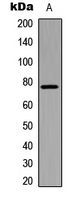
Western blot analysis of BMX (pY566) expression in HeLa (A) whole cell lysates.Application
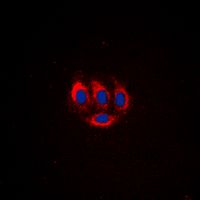
Immunofluorescent analysis of BMX (pY566) staining in HeLa cells. Formalin-fixed cells were permeabilized with 0.1% Triton X-100 in TBS for 5-10 minutes and blocked with 3% BSA-PBS for 30 minutes at room temperature. Cells were probed with the primary antibody in 3% BSA-PBS and incubated overnight at 4 deg C in a hidified chamber. Cells were washed with PBST and incubated with a DyLight 594-conjugated secondary antibody (red) in PBS at room temperature in the dark. DAPI was used to stain the cell nuclei (blue).Application
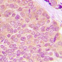
Immunohistochemical analysis of BMX (pY566) staining in human breast cancer formalin fixed paraffin embedded tissue section. The section was pre-treated using heat mediated antigen retrieval with sodium citrate buffer (pH 6.0). The section was then incubated with the antibody at room temperature and detected using an HRP conjugad compact polymer system. DAB was used as the chromogen. The section was then counterstained with haematoxylin and mounted with DPX.| Product Name | BMX (phospho Tyr566) antibody |
|---|---|
| Antibody Type | Primary Antibodies |
| Product description | Rabbit Polyclonal antibody to BMX (phospho Tyr566)This gene encodes a non-receptor tyrosine kinase belonging to the Tec kinase family. The protein contains a PH-like domain, which mediates membrane targeting by binding to phosphatidylinositol 3,4,5-triphosphate (PIP3), and a SH2 domain that binds to tyrosine-phosphorylated proteins and functions in signal transduction. The protein is implicated in several signal transduction pathways including the Stat pathway, and regulates differentiation and tumorigenicity of several types of cancer cells. Multiple alternatively spliced variants, encoding the same protein, have been identified.[provided by RefSeq, Sep 2009] |
| Immunogen | KLH-conjugated synthetic peptide encompassing a sequence within the C-term region of human BMX (phospho Tyr566). The exact sequence is proprietary. |
| Modification | p-Tyr566 |
| Clonality | Polyclonal |
|---|---|
| Isotype | IgG |
| Host Species | Rabbit |
| Tested Applications | ICC/IFIHC-PWB |
| WB: 1:500 - 1:1000. IHC-P: 1:100 - 1:200. ICC/IF: 1:100 - 1:500: |
|
| Species Reactivity | Human |
| Concentration | 1mg/ml |
| Purification | Affinity purified |
| Gene Synonyms | PSCTK3 ETK BMX PSCTK2 BMX non-receptor tyrosine kinase |
|---|---|
| Target | BMX (phospho Tyr566) |
| Tissue Specificity | Recognizes endogenous levels of BMX (pY566) protein. |
Application
Western blot analysis of BMX (pY566) expression in HeLa (A) whole cell lysates.
Application
Immunofluorescent analysis of BMX (pY566) staining in HeLa cells. Formalin-fixed cells were permeabilized with 0.1% Triton X-100 in TBS for 5-10 minutes and blocked with 3% BSA-PBS for 30 minutes at room temperature. Cells were probed with the primary antibody in 3% BSA-PBS and incubated overnight at 4 deg C in a hidified chamber. Cells were washed with PBST and incubated with a DyLight 594-conjugated secondary antibody (red) in PBS at room temperature in the dark. DAPI was used to stain the cell nuclei (blue).
Application
Immunohistochemical analysis of BMX (pY566) staining in human breast cancer formalin fixed paraffin embedded tissue section. The section was pre-treated using heat mediated antigen retrieval with sodium citrate buffer (pH 6.0). The section was then incubated with the antibody at room temperature and detected using an HRP conjugad compact polymer system. DAB was used as the chromogen. The section was then counterstained with haematoxylin and mounted with DPX.| Application Notes | WB: 1:500 - 1:1000. IHC-P: 1:100 - 1:200. ICC/IF: 1:100 - 1:500: |
|---|
| Form | Liquid |
|---|---|
| Storage Instructions | Shipped at 4 °C. Upon delivery aliquot and store at -20 °C for one year. Avoid freeze/thaw cycles. |
| Storage Buffer | Liquid in 0.42% Potassium phosphate; 0.87% Sodium chloride; pH 7.3; 30% glycerol; and 0.01% sodium azide. |


