Application
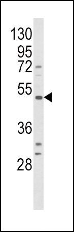
Western blot analysis of BDKRB1 Antibody (Center) in HepG2 cell line lysates (35ug/lane). BDKRB1 (arrow) was detected using the purified Pab.Application
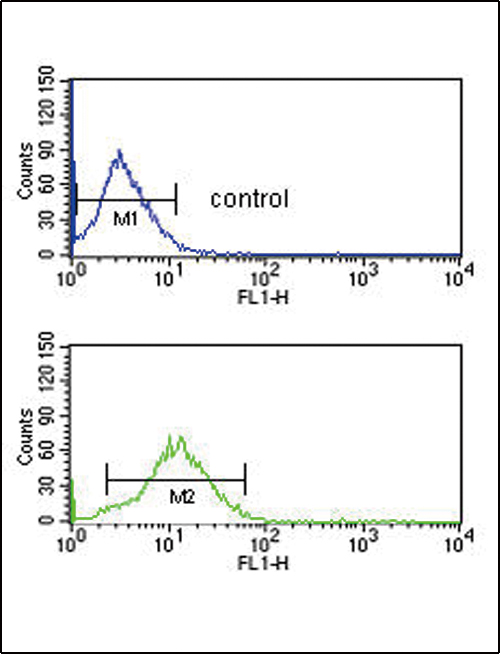
BDKRB1 Antibody (Center) flow cytometric analysis of HepG2 cells (bottom histogram) compared to a negative control cell (top histogram).FITC-conjugated goat-anti-rabbit secondary antibodies were used for the analysis.| Product Name | Bradykinin B1 receptor antibody, Internal |
|---|---|
| Antibody Type | Primary Antibodies |
| Product description | Rabbit polyclonal to Bradykinin B1 receptorBradykinin, a 9 aa peptide, is generated in pathophysiologic conditions such as inflammation, trauma, burns, shock, and allergy. Two types of G-protein coupled receptors have been found which bind bradykinin and mediate responses to these pathophysiologic conditions. The protein encoded by this gene is one of these receptors and is synthesized de novo following tissue injury. Receptor binding leads to an increase in the cytosolic calcium ion concentration, ultimately resulting in chronic and acute inflammatory responses. [provided by RefSeq] |
| Immunogen | KLH conjugated synthetic peptide between 219~249 amino acids from the Center region of human BDKRB1. |
| Clonality | Polyclonal |
|---|---|
| Isotype | IgG |
| Host Species | Rabbit |
| Tested Applications | FACSWB |
| ELISA: Use at 1:1,000: |
|
| Species Reactivity | Human |
| Concentration | 0.2mg/ml |
| Purification | Affinity purified |
| Gene Synonyms | BDKRB1 BRADYB1 B1R B1 bradykinin receptor BK1 receptor B1 bradykinin receptor BK-1 receptor BDKRB1 |
|---|---|
| Target | BDKRB1 |
| Molecular Weight(MW) | 40.495 kDa (note) |
Application
Western blot analysis of BDKRB1 Antibody (Center) in HepG2 cell line lysates (35ug/lane). BDKRB1 (arrow) was detected using the purified Pab.
Application
BDKRB1 Antibody (Center) flow cytometric analysis of HepG2 cells (bottom histogram) compared to a negative control cell (top histogram).FITC-conjugated goat-anti-rabbit secondary antibodies were used for the analysis.| Application Notes | ELISA: Use at 1:1,000: |
|---|
| Form | Liquid |
|---|---|
| Storage Instructions | Store at at 2-8°C for up to 6 months. For long term storage, aliquot and store at -20 °C. Avoid repeated freeze/thaw cycles. |
| Storage Buffer | PBS with 0.09% (W/V) sodium azide |


