Application
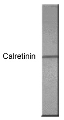
Western blot using anti-Calretinin antibody & calretinin WB positive control.| Product Name | Calretinin antibody |
|---|---|
| Antibody Type | Primary Antibodies |
| Product description | Rabbit polyclonal to CalretininCalretinin is a 29 kDa calcium binding protein that is expressed in central and peripheral nervous system and in many normal and pathological tissues. Calretinin is also considered an important diagnostic aid in the differential diagnosis of cystic and solid ameloblastic tumors. The rat and human calretinin exhibit 98% sequence homology and 91% homology to many other species. Calretinin is generally expressed with two other calcium binding proteins Calbindin D-29 and parvalbumin in CNS and other peripheral tissues. Two calcium-binding proteins, calbindin and calretinin, have been reported to be expressed in abundance in Purkinje cells and other cell types in the cerebellum. The antibodies against calretinin also labels the mossy fibers throughout, and climbing fibers in a distinct group of folia. They also stain several cell types in the molecular layer. There is no appreciable immunostaining of calretinin in cerebellar cortex. In the rat, antibodies against calretinin stain the Lugaro cells, and some granule cells in lobe X; they also give weak staining of all the granule cells in the other lobes. Calretinin antibodies have been used to detect a number of carcinomas by clinicopathological, immunohistochemical techniques including, epithelial malignant mesotheliomas, Ovarian yolk sac tumor, Adenomatoid tumors of the uterus etc. |
| Immunogen | Synthetic cyclic peptide (common to all Calretinin proteins) (Human). |
| Clonality | Polyclonal |
|---|---|
| Isotype | IgG |
| Host Species | Rabbit |
| Tested Applications | IPWB |
| IP: Use at a dilution of 1/250. WB: Use at a dilution of 1/1000. Detects a band of approximately 29 kDa. Optimal dilutions/concentrations should be determined by the end user.: |
|
| Species Reactivity | HumanMouseRat |
| Concentration | 1mg/ml (Please refer to the vial label for the specific concentration) |
| Purification | Affinity purified |
| Gene Synonyms | Calretinin 114051 794 CAL2 CALB2 P22676 |
|---|---|
| Target | Calretinin |
| Tissue Specificity | Has no affinity for other calcium binding proteins including Calbindin, parvalbumin, and S100 protein. |
Application
Western blot using anti-Calretinin antibody & calretinin WB positive control.| Positive Control | Rat brain homogenates. |
|---|---|
| Application Notes | IP: Use at a dilution of 1/250. WB: Use at a dilution of 1/1000. Detects a band of approximately 29 kDa. Optimal dilutions/concentrations should be determined by the end user.: |
| Form | Liquid |
|---|---|
| Storage Instructions | Keep as concentrated solution. Store at 4ºC short term. For extended storage aliquot and store at -20ºC or below. Avoid freeze-thaw cycles. |
| Storage Buffer | Preservative: 0.02% Thimerosal / Merthiolate |


