Application
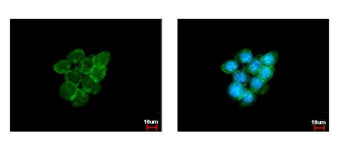
Caveolin 1 antibody [N1N3] detects Caveolin 1 protein at membrane by immunofluorescent analysis. Sample: A431 cells were fixed in ice-cold MeOH for 5 min. Green: Caveolin 1 protein stained by Caveolin 1 antibody [N1N3] diluted at 1:500. Blue: Hoechst 33343 staining.Application
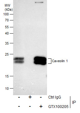
Immunoprecipitation of Caveolin 1 protein from A549 membrane extracts using 5 μg of Caveolin 1 antibody [N1N3] . Western blot analysis was performed using Caveolin 1 antibody [N1N3] . EasyBlot anti-Rabbit IgG was used as a secondary reagent.Application
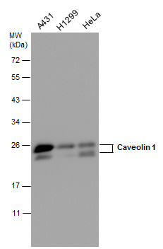
Caveolin 1 antibody detects Caveolin 1 protein by western blot analysis. Various whole cell extracts (30 µg) were separated by 12% SDS-PAGE, and the membrane was blotted with Caveolin 1 antibody diluted at a dilution of 1:500. The HRP-conjugated anti-rabbit IgG antibody was used to detect the primary antibody.Application
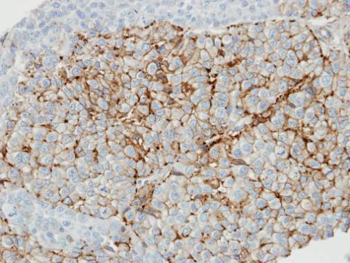
Immunohistochemical microscopy analysis of paraffin-embedded lung carcinoma cell line xenograft tissue using Caveolin-1 antibody [N1N3] (1:100).Application
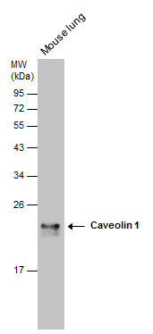
Caveolin 1 antibody [N1N3] detects Caveolin 1 protein by western blot analysis. Mouse tissue extracts (50 µg) was separated by 12% SDS-PAGE, and the membrane was blotted with Caveolin 1 antibody [N1N3] diluted at 1:500. The HRP-conjugated anti-rabbit IgG antibody was used to detect the primary antibody.| Product Name | Caveolin 1 antibody |
|---|---|
| Antibody Type | Primary Antibodies |
| Product description | Rabbit polyclonal antibody to Caveolin 1 (caveolin 1, caveolae protein, 22kDa)The scaffolding protein encoded by this gene is the main component of the caveolae plasma membranes found in most cell types. The protein links integrin subunits to the tyrosine kinase FYN, an initiating step in coupling integrins to the Ras-ERK pathway and promoting cell cycle progression. The gene is a tumor suppressor gene candidate and a negative regulator of the Ras-p42/44 MAP kinase cascade. CAV1 and CAV2 are located next to each other on chromosome 7 and express colocalizing proteins that form a stable hetero-oligomeric complex. By using alternative initiation codons in the same reading frame, two isoforms (alpha and beta) are encoded by a single transcript from this gene. [provided by RefSeq] |
| Immunogen | Carrier-protein conjugated synthetic peptide encompassing a sequence within the N-terminus region of human Caveolin 1. The exact sequence is proprietary. |
| Clonality | Polyclonal |
|---|---|
| Isotype | IgG |
| Host Species | Rabbit |
| Tested Applications | ICC/IFIHCIHC-PIPWB |
| *Optimal dilutions/concentrations should be determined by the researcher.
: |
|
| Species Reactivity | HumanMouseRat |
| Predicted Reactivity | Rhesus Monkey, ChimpanzeeRhesus Monkey (100%), Chimpanzee (100%) |
| Concentration | 1mg/ml (Please refer to the vial label for the specific concentration) |
| Purification | Affinity purified |
| Gene Synonyms | BSCL3 antibody CGL3 antibody MSTP085 antibody VIP21 antibody CAV1 antibody cell growth-inhibiting protein 32 antibody caveolin-1 antibody "caveolin 1 caveolae protein 22kDa antibody" |
|---|---|
| Target | Caveolin 1 |
| Molecular Weight(MW) | 20 kDa (note) (4) |
| Cellular Localization | Golgi apparatus membrane , Cell membrane , Membrane , caveola |
Application
Caveolin 1 antibody [N1N3] detects Caveolin 1 protein at membrane by immunofluorescent analysis. Sample: A431 cells were fixed in ice-cold MeOH for 5 min. Green: Caveolin 1 protein stained by Caveolin 1 antibody [N1N3] diluted at 1:500. Blue: Hoechst 33343 staining.
Application
Immunoprecipitation of Caveolin 1 protein from A549 membrane extracts using 5 μg of Caveolin 1 antibody [N1N3] . Western blot analysis was performed using Caveolin 1 antibody [N1N3] . EasyBlot anti-Rabbit IgG was used as a secondary reagent.
Application
Caveolin 1 antibody detects Caveolin 1 protein by western blot analysis. Various whole cell extracts (30 µg) were separated by 12% SDS-PAGE, and the membrane was blotted with Caveolin 1 antibody diluted at a dilution of 1:500. The HRP-conjugated anti-rabbit IgG antibody was used to detect the primary antibody.
Application
Immunohistochemical microscopy analysis of paraffin-embedded lung carcinoma cell line xenograft tissue using Caveolin-1 antibody [N1N3] (1:100).
Application
Caveolin 1 antibody [N1N3] detects Caveolin 1 protein by western blot analysis. Mouse tissue extracts (50 µg) was separated by 12% SDS-PAGE, and the membrane was blotted with Caveolin 1 antibody [N1N3] diluted at 1:500. The HRP-conjugated anti-rabbit IgG antibody was used to detect the primary antibody.| Positive Control | A431 , H1299 , HeLa , *A549 , *A7 , Mouse lung |
|---|---|
| Application Notes | *Optimal dilutions/concentrations should be determined by the researcher.
: |
| Form | Liquid |
|---|---|
| Storage Instructions | Keep as concentrated solution. Aliquot and store at -20ºC or below. Avoid multiple freeze-thaw cycles. |
| Storage Buffer | 1XPBS, 1% BSA, 20% Glycerol (pH7). 0.01% Thimerosal was added as a preservative. |


