Application
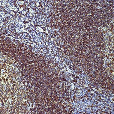
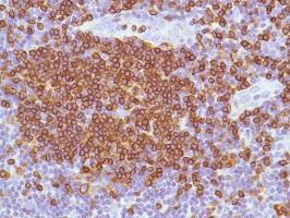
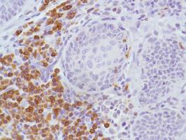
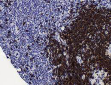
Formalin fixed paraffin embedded human tonsil stained with CD79a antibody (cat. # ).Application
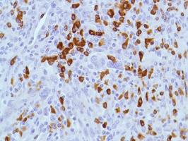
Human HK Lymphoma stained with anti-CD79a antibodyApplication
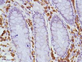
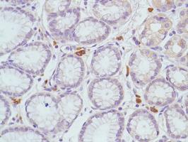
Human Colon stained with anti-CD79a antibodyApplication
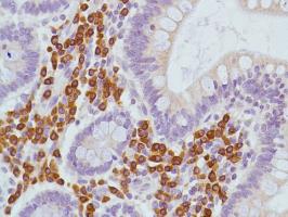
Human Small Intestine stained with anti-CD79a antibodyApplication
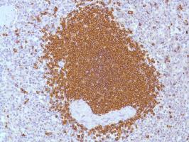
Human Reactive Lymph Node stained with anti-CD79a antibodyApplication
Human Spleen stained with anti-CD79a antibodyApplication
Human Stomach stained with anti-CD79a antibodyApplication
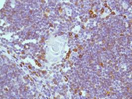
Human Thymus stained with anti-CD79a antibodyApplication
Human Cervial Squamous Cell Carcinoma stained with anti-CD79a antibodyApplication
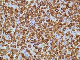
Human B Cell Lymphoma stained with anti-CD79a antibodyApplication
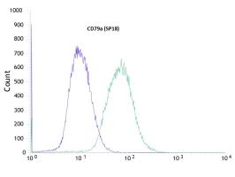
Flow cytometric analysis of rabbit anti-CD79a (SP18) antibody in Jurkat (green) compare to negative control of rabbit IgG (blue)Application
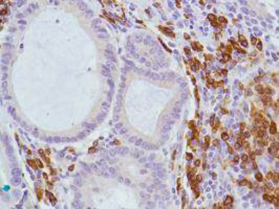
Human stomach adenocarcinoma stained with anti-CD79a antibodyApplication
Human B Cell Lymphoma stained with anti-CD79a antibody| Product Name | CD79a antibody |
|---|---|
| Antibody Type | Primary Antibodies |
| Product description | Monoclonal Rabbit Antibody to Human CD79aThe B lymphocyte antigen receptor is a multimeric complex that includes the antigen-specific component, surface immunoglobulin (Ig). Surface Ig non-covalently associates with two other proteins, Ig-alpha and Ig-beta, which are necessary for expression and function of the B-cell antigen receptor. This gene encodes the Ig-alpha protein of the B-cell antigen component. Alternatively spliced transcript variants encoding different isoforms have been described. [provided by RefSeq] |
| Immunogen | Synthetic peptide derived from N-terminal region of human CD79a protein. |
| Clonality | Monoclonal |
|---|---|
| Isotype | IgG |
| Clone Number | SP18 |
| Host Species | Rabbit |
| Tested Applications | FACSIHCIHC-P |
| We suggest an incubation period of 30 minutes at room temperature. However, depending upon the fixation conditions and the staining system employed, optimal incubation should be determined by the user. Formalin fixed paraffin embedded tissue sections require high temperature antigen unmasking with 10 mM citrate buffer, pH 6.0 prior to immunostaining.: |
|
| Species Reactivity | Human |
| Concentration | 1mg/ml |
| Gene Synonyms | 112205 973 IGA P11912 MB-1 CD79A MB1 MB 1 |
|---|---|
| Target | CD79a |
| Tissue Specificity | This antibody recognizes CD79a. CD79a is a disulphide-linked heterodimer, consisting of CD79a / mb-1 and CD79b / B29 polypeptides, is non-covalently associated with membrane-bound immunoglobulins on B cells to constitute the B cell Ag receptor. CD79a first appears at pre B cell stage and persists until the plasma cell stage where it is found as an intracellular component. CD79a is found in the majority of acute leukemias of precursor B cell type, in B cell lines, B cell lymphomas, and in some myelomas. This antibody crossreacts with Human. |
| Cellular Localization | Cell membrane |
Application
Formalin fixed paraffin embedded human tonsil stained with CD79a antibody (cat. # ).
Application
Human HK Lymphoma stained with anti-CD79a antibody
Application
Human Colon stained with anti-CD79a antibody
Application
Human Small Intestine stained with anti-CD79a antibody
Application
Human Reactive Lymph Node stained with anti-CD79a antibody
Application
Human Spleen stained with anti-CD79a antibody
Application
Human Stomach stained with anti-CD79a antibody
Application
Human Thymus stained with anti-CD79a antibody
Application
Human Cervial Squamous Cell Carcinoma stained with anti-CD79a antibody
Application
Human B Cell Lymphoma stained with anti-CD79a antibody
Application
Flow cytometric analysis of rabbit anti-CD79a (SP18) antibody in Jurkat (green) compare to negative control of rabbit IgG (blue)
Application
Human stomach adenocarcinoma stained with anti-CD79a antibody
Application
Human B Cell Lymphoma stained with anti-CD79a antibody| Positive Control | Tonsil |
|---|---|
| Application Notes | We suggest an incubation period of 30 minutes at room temperature. However, depending upon the fixation conditions and the staining system employed, optimal incubation should be determined by the user. Formalin fixed paraffin embedded tissue sections require high temperature antigen unmasking with 10 mM citrate buffer, pH 6.0 prior to immunostaining.: |
| Form | Liquid |
|---|---|
| Storage Instructions | Keep as concentrated solution, aliquot and store at 4°C. Do not freeze. |
| Storage Buffer | This antibody is supplied as tissue culture supernatant containing sodium azide as preservative. |


