Application
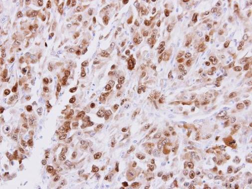
Immunohistochemical analysis of paraffin-embedded ES2 xenograft, using CDT1 antibody at 1:500 dilution.Application
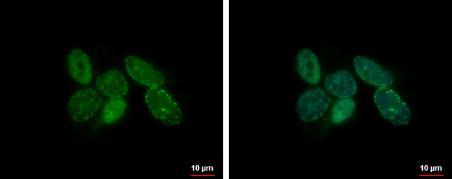
CDT1 antibody detects CDT1 protein at nucleus by immunofluorescent analysis. Sample: SK-N-AS cells were fixed in 4% paraformaldehyde at RT for 15 min. Green: CDT1 protein stained by CDT1 antibody diluted at 1:500. Blue: Hoechst 33342 staining.Application
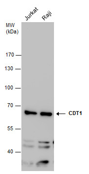
CDT1 antibody detects CDT1 protein by western blot analysis. Various whole cell extracts (30 μg) were separated by 7.5% SDS-PAGE, and the membrane was blotted with CDT1 antibody diluted at a dilution of 1:1000.| Product Name | CDT1 antibody |
|---|---|
| Antibody Type | Primary Antibodies |
| Product description | Rabbit Polyclonal antibody to CDT1 (chromatin licensing and DNA replication factor 1)Cooperates with CDC6 to promote the loading of the mini-chromosome maintenance complex onto chromatin to form the pre-replication complex necessary to initiate DNA replication. Binds DNA in a sequence-, strand-, and conformation-independent manner. Potential oncogene. |
| Clonality | Polyclonal |
|---|---|
| Isotype | IgG |
| Host Species | Rabbit |
| Tested Applications | ICC/IFIHC-PWB |
| *Optimal dilutions/concentrations should be determined by the researcher.
: |
|
| Species Reactivity | Human |
| Concentration | 1mg/ml |
| Purification | Affinity purified |
| Gene Synonyms | DUP antibody RIS2 antibody CDT1 antibody "Double parked Drosophila homolog of antibody" DNA replication factor Cdt1 antibody chromatin licensing and DNA replication factor 1 antibody |
|---|---|
| Molecular Weight(MW) | 60 kDa (note) |
| Cellular Localization | Nucleus |
Application
Immunohistochemical analysis of paraffin-embedded ES2 xenograft, using CDT1 antibody at 1:500 dilution.
Application
CDT1 antibody detects CDT1 protein at nucleus by immunofluorescent analysis. Sample: SK-N-AS cells were fixed in 4% paraformaldehyde at RT for 15 min. Green: CDT1 protein stained by CDT1 antibody diluted at 1:500. Blue: Hoechst 33342 staining.
Application
CDT1 antibody detects CDT1 protein by western blot analysis. Various whole cell extracts (30 μg) were separated by 7.5% SDS-PAGE, and the membrane was blotted with CDT1 antibody diluted at a dilution of 1:1000.| Positive Control | Jurkat , Raji |
|---|---|
| Application Notes | *Optimal dilutions/concentrations should be determined by the researcher.
: |
| Form | Liquid |
|---|---|
| Storage Instructions | Keep as concentrated solution. Aliquot and store at -20ºC or below. Avoid multiple freeze-thaw cycles. |
| Storage Buffer | 1XPBS, 20% Glycerol (pH7). 0.025% ProClin 300 was added as a preservative. |


