Application
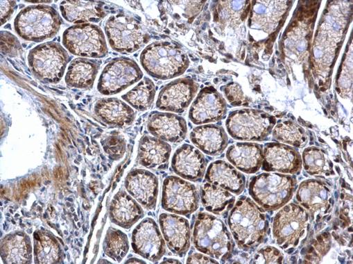
Chk1 (phospho Ser345) antibody [C1C2], Internal detects Chk1 (phospho Ser345) protein at nucleus on mouse colon by immunohistochemical analysis. Sample: Paraffin-embedded mouse colon. Chk1 (phospho Ser345) antibody [C1C2], Internal dilution: 1:500.Application
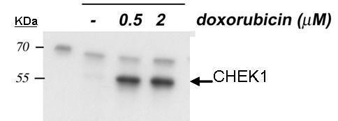
Sample: 20 µg of HCT116 P53 whole cell lysate 10% SDS PAGE Chk1-phopho-S345 antibody diluted at 1:1000 The HRP-conjugated anti-rabbit IgG antibody was used to detect the primary antibody.Application
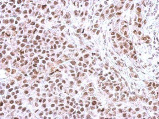
Immunohistochemical analysis of paraffin-embedded HeLa xenograft, using Chk1 (phospho Ser345) antibody at 1:500 dilution.Application
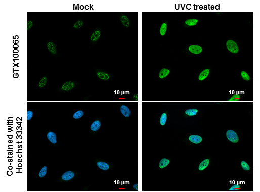
Chk1 (phospho Ser345) antibody [C1C2], Internal detects Chk1 (phospho Ser345) protein at nucleus by immunofluorescent analysis. Samples: HeLa cells mock (left) and treated with 100 J/m2 UVC and recover for 8 hrs (right) were fixed in 4% paraformaldehyde at RT for 15 min. Green: Chk1 (phospho Ser345) protein stained by Chk1 (phospho Ser345) antibody [C1C2], Internal diluted at 1:500. Blue: Hoechst 33342 staining. Scale bar = 10 μm.Application
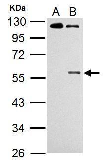
Sample (30 µg of whole cell lysate) A: HCT116 cells with mock treatment for 24 hr B: HCT116 cells with 30 µM cisplatin treatment for 24 hr 10% SDS PAGE diluted at 1:500 The HRP-conjugated anti-rabbit IgG antibody was used to detect the primary antibody.| Product Name | Chk1 (phospho Ser345) antibody [C1C2], Internal |
|---|---|
| Antibody Type | Primary Antibodies |
| Product description | Rabbit Polyclonal antibody to Chk1 (phospho Ser345) (CHK1 checkpoint homolog (S. pombe))Required for checkpoint mediated cell cycle arrest in response to DNA damage or the presence of unreplicated DNA. May also negatively regulate cell cycle progression during unperturbed cell cycles. Recognizes the substrate consensus sequence [R-X-X-S/T]. Binds to and phosphorylates CDC25A, CDC25B and CDC25C. Phosphorylation of CDC25A at 'Ser-178' and 'Thr-507' and phosphorylation of CDC25C at 'Ser-216' creates binding sites for 14-3-3 proteins which inhibit CDC25A and CDC25C. Phosphorylation of CDC25A at 'Ser-76', 'Ser-124', 'Ser-178', 'Ser-279' and 'Ser-293' promotes proteolysis of CDC25A. Inhibition of CDC25 activity leads to increased inhibitory tyrosine phosphorylation of CDK-cyclin complexes and blocks cell cycle progression. Binds to and phosphorylates RAD51 at 'Thr-309', which may enhance the association of RAD51 with chromatin and promote DNA repair by homologous recombination. Binds to and phosphorylates TLK1 at 'Ser-743', which prevents the TLK1-dependent phosphorylation of the chromatin assembly factor ASF1A. This may affect chromatin assembly during S phase or DNA repair. May also phosphorylate multiple sites within the C-terminus of TP53, which promotes activation of TP53 by acetylation and enhances suppression of cellular proliferation. |
| Modification | p-Ser345 |
| Clonality | Polyclonal |
|---|---|
| Isotype | IgG |
| Host Species | Rabbit |
| Tested Applications | ICC/IFIHC-PWB |
| *Optimal dilutions/concentrations should be determined by the researcher. | |
| Species Reactivity | HumanMouse |
| Predicted Reactivity | Rat, BovineRat (100%), Bovine (100%) |
| Concentration | 1mg/ml (Please refer to the vial label for the specific concentration) |
| Purification | Affinity purified |
| Gene Synonyms | CHK1 antibody, CHEK1 antibody, cell cycle checkpoint kinase antibody, "Checkpoint, S. pombe, homolog of, 1 antibody", serine/threonine-protein kinase Chk1 antibody, checkpoint kinase-1 antibody, CHK1 checkpoint homolog antibody, CHK1 checkpoint homo |
|---|---|
| Molecular Weight(MW) | 54 kDa (note) (4) |
| Cellular Localization | Nucleus , Cytoplasm |
Application
Chk1 (phospho Ser345) antibody [C1C2], Internal detects Chk1 (phospho Ser345) protein at nucleus on mouse colon by immunohistochemical analysis. Sample: Paraffin-embedded mouse colon. Chk1 (phospho Ser345) antibody [C1C2], Internal dilution: 1:500.
Application
Sample: 20 µg of HCT116 P53 whole cell lysate 10% SDS PAGE Chk1-phopho-S345 antibody diluted at 1:1000 The HRP-conjugated anti-rabbit IgG antibody was used to detect the primary antibody.
Application
Immunohistochemical analysis of paraffin-embedded HeLa xenograft, using Chk1 (phospho Ser345) antibody at 1:500 dilution.
Application
Chk1 (phospho Ser345) antibody [C1C2], Internal detects Chk1 (phospho Ser345) protein at nucleus by immunofluorescent analysis. Samples: HeLa cells mock (left) and treated with 100 J/m2 UVC and recover for 8 hrs (right) were fixed in 4% paraformaldehyde at RT for 15 min. Green: Chk1 (phospho Ser345) protein stained by Chk1 (phospho Ser345) antibody [C1C2], Internal diluted at 1:500. Blue: Hoechst 33342 staining. Scale bar = 10 μm.
Application
Sample (30 µg of whole cell lysate) A: HCT116 cells with mock treatment for 24 hr B: HCT116 cells with 30 µM cisplatin treatment for 24 hr 10% SDS PAGE diluted at 1:500 The HRP-conjugated anti-rabbit IgG antibody was used to detect the primary antibody.| Positive Control | HCT116 , HCT116 cells with 30uM cisplatin treatment for 24hr |
|---|---|
| Application Notes | *Optimal dilutions/concentrations should be determined by the researcher. |
| Form | Liquid |
|---|---|
| Storage Instructions | Keep as concentrated solution. Aliquot and store at -20ºC or below. Avoid multiple freeze-thaw cycles. |
| Storage Buffer | 1XPBS, 1% BSA, 20% Glycerol (pH7). 0.01% Thimerosal was added as a preservative. |


