Application
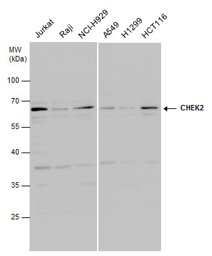
WB ImageCHEK2 antibody detects CHEK2 protein by western blot analysis. Various whole cell extracts (30 μg) were separated by 10% SDS-PAGE, and the membrane was blotted with CHEK2 antibody diluted at 1:3000.| Product Name | Chk2 antibody |
|---|---|
| Antibody Type | Primary Antibodies |
| Product description | Rabbit Polyclonal antibody to Chk2 (CHK2 checkpoint homolog (S. pombe))In response to DNA damage and replication blocks, cell cycle progression is halted through the control of critical cell cycle regulators. The protein encoded by this gene is a cell cycle checkpoint regulator and putative tumor suppressor. It contains a forkhead-associated protein interaction domain essential for activation in response to DNA damage and is rapidly phosphorylated in response to replication blocks and DNA damage. When activated, the encoded protein is known to inhibit CDC25C phosphatase, preventing entry into mitosis, and has been shown to stabilize the tumor suppressor protein p53, leading to cell cycle arrest in G1. In addition, this protein interacts with and phosphorylates BRCA1, allowing BRCA1 to restore survival after DNA damage. Mutations in this gene have been linked with Li-Fraumeni syndrome, a highly penetrant familial cancer phenotype usually associated with inherited mutations in TP53. Also, mutations in this gene are thought to confer a predisposition to sarcomas, breast cancer, and brain tumors. This nuclear protein is a member of the CDS1 subfamily of serine/threonine protein kinases. Three transcript variants encoding different isoforms have been found for this gene. [provided by RefSeq] |
| Immunogen | Recombinant protein encompassing a sequence within the center region of human Chk2. The exact sequence is proprietary. |
| Clonality | Polyclonal |
|---|---|
| Isotype | IgG |
| Host Species | Rabbit |
| Tested Applications | WB |
| *Optimal dilutions/concentrations should be determined by the researcher. | |
| Species Reactivity | Human |
| Predicted Reactivity | Mouse, Rat, Pig, BovineMouse (91%), Rat (91%), Pig (93%), Bovine (94%) |
| Concentration | 1mg/ml (Please refer to the vial label for the specific concentration) |
| Purification | Affinity purified |
| Gene Synonyms | CDS1 antibody, CHK2 antibody, HuCds1 antibody, LFS2 antibody, PP1425 antibody, RAD53 antibody, CHEK2 antibody, protein kinase CHK2 antibody, checkpoint-like protein CHK2 antibody, cds1 homolog antibody, serine/threonine-protein kinase Chk2 antibody, CHK2 c |
|---|---|
| Molecular Weight(MW) | 61 kDa (note) (4) |
| Cellular Localization | Nucleoplasm , Nucleus , PML body |
Application
WB ImageCHEK2 antibody detects CHEK2 protein by western blot analysis. Various whole cell extracts (30 μg) were separated by 10% SDS-PAGE, and the membrane was blotted with CHEK2 antibody diluted at 1:3000.| Positive Control | Jurkat , Raji , NCI-H929 , A549 , H1299 , HCT116 |
|---|---|
| Application Notes | *Optimal dilutions/concentrations should be determined by the researcher. |
| Form | Liquid |
|---|---|
| Storage Instructions | Keep as concentrated solution. Aliquot and store at -20ºC or below. Avoid multiple freeze-thaw cycles. |
| Storage Buffer | 1XPBS, 1% BSA, 20% Glycerol (pH7). 0.01% Thimerosal was added as a preservative. |


