Application
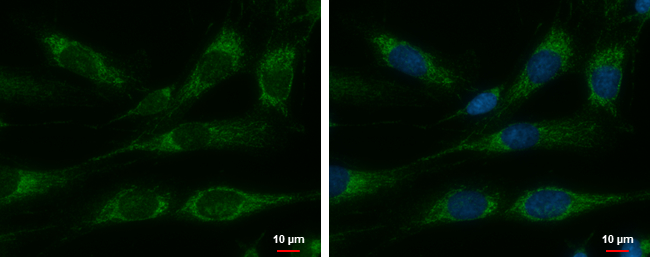
Cpn10 antibody detects Cpn10 protein at mitochondria by immunofluorescent analysis. Sample: SKNSH cells were fixed in 2% paraformaldehyde/culture medium at 37℃ for 15 min. Green: Cpn10 protein stained by Cpn10 antibody diluted at 1:500. Blue: Hoechst 33342 staining.Application
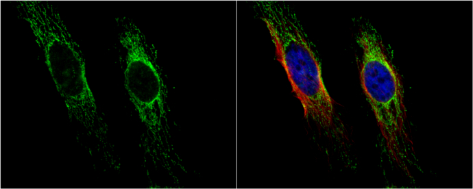
Cpn10 antibody detects Cpn10 protein at mitochondria by immunofluorescent analysis. Sample: HeLa cells were fixed in 4% paraformaldehyde at RT for 15 min. Green: Cpn10 protein stained by Cpn10 antibody diluted at 1:100. Red: alpha Tubulin, a cytoskeleton marker, stained by alpha Tubulin antibody [B-5-1-2] diluted at 1:10000. Blue: Hoechst 33342 staining.Application
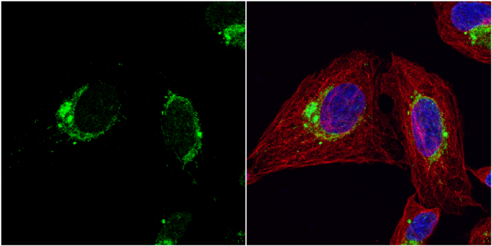
Cpn10 antibody detects Cpn10 protein at cytoplasm by immunofluorescent analysis. Sample: U2OS cells were fixed in 4% paraformaldehyde at RT for 15 min. Green: Cpn10 protein stained by Cpn10 antibody diluted at 1:200. Red: alpha Tubulin, a cytoskeleton marker, stained by alpha Tubulin antibody [GT114] diluted at 1:1000. Blue: Hoechst 33342 staining.Application
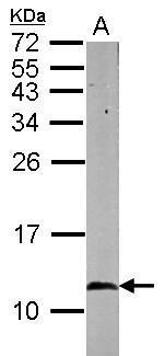
Sample (50 ug of whole cell lysate) A: mouse brain 15% SDS PAGE diluted at 1:1000Application
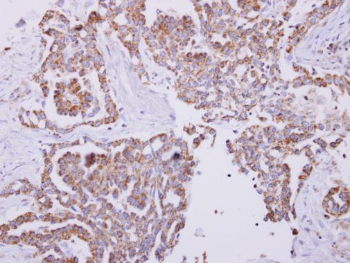
Cpn10 antibody detects Cpn10 protein at cytoplasm on human lung carcinoma by immunohistochemical analysis. Sample: Paraffin-embedded lung carcinoma. Cpn10 antibody dilution: 1:250.| Product Name | Cpn10 antibody |
|---|---|
| Antibody Type | Primary Antibodies |
| Product description | Rabbit Polyclonal antibody to Cpn10 (heat shock 10kDa protein 1 (chaperonin 10))This gene encodes a major heat shock protein which functions as a chaperonin. Its structure consists of a heptameric ring which binds to another heat shock protein in order to form a symmetric, functional heterodimer which enhances protein folding in an ATP-dependent manner. This gene and its co-chaperonin, HSPD1, are arranged in a head-to-head orientation on chromosome 2. [provided by RefSeq] |
| Immunogen | Full length human Cpn10 Recombinant protein. |
| Clonality | Polyclonal |
|---|---|
| Isotype | IgG |
| Host Species | Rabbit |
| Tested Applications | ICC/IFIHC-PWB |
| *Optimal dilutions/concentrations should be determined by the researcher. | |
| Species Reactivity | HumanMouse |
| Predicted Reactivity | Rat, Zebrafish, Japanese medaka, Xenopus laevis, Pig, Chicken, Rhesus Monkey, BovineRat (98%), Zebrafish (80%), Japanese medaka (83%), Xenopus laevis (88%), Pig (100%), Chicken (87%), Rhesus Monkey (99%), Bovine (100%) |
| Concentration | 1mg/ml (Please refer to the vial label for the specific concentration) |
| Purification | Affinity purified |
| Gene Synonyms | CPN10 antibody, EPF antibody, GROES antibody, HSP10 antibody, HSPE1 antibody, "10 kDa heat shock protein, mitochondrial antibody", 10 kDa chaperonin antibody, chaperonin 10 antibody, early-pregnancy factor antibody, heat shock 10kD protein 1 (chaperonin 10 |
|---|---|
| Molecular Weight(MW) | 11 kDa (note) (2) |
| Cellular Localization | Mitochondrion matrix |
Application
Cpn10 antibody detects Cpn10 protein at mitochondria by immunofluorescent analysis. Sample: SKNSH cells were fixed in 2% paraformaldehyde/culture medium at 37℃ for 15 min. Green: Cpn10 protein stained by Cpn10 antibody diluted at 1:500. Blue: Hoechst 33342 staining.
Application
Cpn10 antibody detects Cpn10 protein at mitochondria by immunofluorescent analysis. Sample: HeLa cells were fixed in 4% paraformaldehyde at RT for 15 min. Green: Cpn10 protein stained by Cpn10 antibody diluted at 1:100. Red: alpha Tubulin, a cytoskeleton marker, stained by alpha Tubulin antibody [B-5-1-2] diluted at 1:10000. Blue: Hoechst 33342 staining.
Application
Cpn10 antibody detects Cpn10 protein at cytoplasm by immunofluorescent analysis. Sample: U2OS cells were fixed in 4% paraformaldehyde at RT for 15 min. Green: Cpn10 protein stained by Cpn10 antibody diluted at 1:200. Red: alpha Tubulin, a cytoskeleton marker, stained by alpha Tubulin antibody [GT114] diluted at 1:1000. Blue: Hoechst 33342 staining.
Application
Sample (50 ug of whole cell lysate) A: mouse brain 15% SDS PAGE diluted at 1:1000
Application
Cpn10 antibody detects Cpn10 protein at cytoplasm on human lung carcinoma by immunohistochemical analysis. Sample: Paraffin-embedded lung carcinoma. Cpn10 antibody dilution: 1:250.| Application Notes | *Optimal dilutions/concentrations should be determined by the researcher. |
|---|
| Form | Liquid |
|---|---|
| Storage Instructions | Keep as concentrated solution. Aliquot and store at -20ºC or below. Avoid multiple freeze-thaw cycles. |
| Storage Buffer | 0.1M Tris, 0.1M Glycine, 20% Glycerol (pH7). 0.01% Thimerosal was added as a preservative. |


