Application
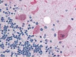
GeneTex anti-Cyclin B1 antibody was diluted 1:500 to detect Cyclin B1 in human brain cerebellum tissue. Tissue was formalin fixed and paraffin embedded. No pre-treatment of sample was required. The image shows the localization of antibody as the precipitated red signal, with a hematoxylin purple nuclear counter stain.Application
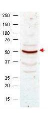
Western blot analysis using GeneTex Anti-Cyclin B1 antibody shows detection of Cyclin B1 present in asynchronous HeLa cell lysates. Comparison to a molecular weight marker indicates a band of ~55 kDa corresponding to human Cyclin B1 (arrowhead). Approximately 50 µg of lysate was loaded on to a 7% SDS-PAGE gel for separation. After transfer to nitrocellulose, the blot was incubated with a 1:500 dilution of the antibody for 1 h at room temperature. Detection occurred using a 1:10,000 of HRP conjugated Goat anti Rabbit IgG .Application
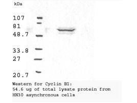
Western blot analysis using GeneTex Anti-Cyclin B1 antibody shows detection of human Cyclin B1 present in asynchronous HN30 cell lysates. HN30 cells are from head and neck cancer tumors that over express cyclin B1 and D1. Comparison to a molecular weight marker indicates a band of ~62 kDa corresponding to the expected molecular weight for the protein. The blot was incubated with a 1:500 dilution of the antibody for 1 h at room temperature. Detection occurred using a 1:10,000 of HRP conjugated Goat anti Rabbit IgG and chemiluminescence reagent with a 1min exposure time.| Product Name | Cyclin B1 antibody |
|---|---|
| Antibody Type | Primary Antibodies |
| Product description | Rabbit Polyclonal antibody to Cyclin B1In mammals, cyclin B associates with inactive p34 cdc2 which facilitates phosphorylation of p34 cdc2 at aa 14 Thr and 15 Tyr. This maintains the inactive state until the end of G2 phase. The inactive cyclin B p34 cdc2 complex continues to accumulate in the cytoplasm until the completion of DNA synthesis, when Cdc25, a specific protein phosphatase, dephosphorylates aa 14Thr and 15Tyr of p34 cdc2 rendering the complex active at the G2 / M boundary. This mitotic kinase complex remains active until the metaphase / anaphase transition when cyclin B is degraded. This degradation process is ubiquitin dependent and is necessary for the cell to exit mitosis. Therefore, cyclin B p34 cdc2 plays a critical role in G2 to M transition. |
| Immunogen | Anti-Cyclin B1 antibody was produced by repeated immunizations of full length fusion protein corresponding to the human gene. |
| Clonality | Polyclonal |
|---|---|
| Host Species | Rabbit |
| Tested Applications | ELISAIHC-PIPWB |
| *Optimal dilutions/concentrations should be determined by the researcher.
: |
|
| Species Reactivity | HumanMouseRat |
| Concentration | 1mg/ml |
| Purification | Affinity purified |
| Gene Synonyms | CCNB1 CCNB |
|---|---|
| Target | Cyclin B1 |
Application
GeneTex anti-Cyclin B1 antibody was diluted 1:500 to detect Cyclin B1 in human brain cerebellum tissue. Tissue was formalin fixed and paraffin embedded. No pre-treatment of sample was required. The image shows the localization of antibody as the precipitated red signal, with a hematoxylin purple nuclear counter stain.
Application
Western blot analysis using GeneTex Anti-Cyclin B1 antibody shows detection of Cyclin B1 present in asynchronous HeLa cell lysates. Comparison to a molecular weight marker indicates a band of ~55 kDa corresponding to human Cyclin B1 (arrowhead). Approximately 50 µg of lysate was loaded on to a 7% SDS-PAGE gel for separation. After transfer to nitrocellulose, the blot was incubated with a 1:500 dilution of the antibody for 1 h at room temperature. Detection occurred using a 1:10,000 of HRP conjugated Goat anti Rabbit IgG .
Application
Western blot analysis using GeneTex Anti-Cyclin B1 antibody shows detection of human Cyclin B1 present in asynchronous HN30 cell lysates. HN30 cells are from head and neck cancer tumors that over express cyclin B1 and D1. Comparison to a molecular weight marker indicates a band of ~62 kDa corresponding to the expected molecular weight for the protein. The blot was incubated with a 1:500 dilution of the antibody for 1 h at room temperature. Detection occurred using a 1:10,000 of HRP conjugated Goat anti Rabbit IgG and chemiluminescence reagent with a 1min exposure time.| Positive Control | Total lysate protein from HN30 asynchronous cells |
|---|---|
| Application Notes | *Optimal dilutions/concentrations should be determined by the researcher.
: |
| Form | Liquid |
|---|---|
| Storage Instructions | Store vial at -20° C prior to opening. Aliquot contents and freeze at -20° C or below for extended storage. Avoid cycles of freezing and thawing. |
| Storage Buffer | 0.01% (w/v) Sodium Azide |


