Application
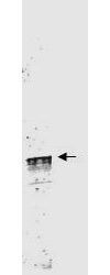
Western blot using GeneTex's Protein A Purified anti-Cyclin L1a antibody shows detection of a band ~59 kDa corresponding to a Cyclin L1a (arrowhead) present in mouse brain whole cell lysate. Approximately 35µg of lysate was separated by 4-20% SDS-PAGE followed by transfer to nitrocellulose. After blocking the membrane was probed with the primary antibody diluted to 1:3,500 for 2h at room temperature.Application
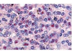
GeneTex's anti-Cyclin L1a antibody was used at a 10 µg/ml to detect Cyclin L1a in a variety of tissues including breast (collagen), heart, kidney (distal tubules), liver, skeletal muscle, ovary µgranulosa and oocyte), pancreas (islet), placenta (trophoblast), prostate (epithelium), skin, spleen (endothelium), stomach (chief), testes (seminiferous epithelium and leydig), thymus (Has-sal's corpuscle and lymphocytes) and uterus µglandular epithelium and stroma). Low to moderate background staining was noted. This image shows Cyclin L1a staining of human spleen. Tissue was formalin-fixed and paraffin embedded.| Product Name | Cyclin L1 (Isoform 1) antibody |
|---|---|
| Antibody Type | Primary Antibodies |
| Product description | Rabbit Polyclonal antibody to Cyclin L1 (Isoform 1)Cyclin L (also referred to as CCNL) is encoded by two highly related genes Cyclin L1 and Cyclin L2 (CCNL1 and CCNL2, respectively). Cyclin L has been shown to associate with the PITSLRE kinase and is involved in pre-mRNA processing. Both Cyclin L gene products can be alternatively spliced to produce two isoforms (known as 1 and 2 or alpha and beta). Cyclin L2 is ubiquitous, expressed at much higher levels than Cyclin L1, and thus is likely the major partner for the CDK11p110 protein kinase. |
| Immunogen | This Protein A purified antibody was prepared from whole rabbit serum produced by repeated immunizations with a synthetic polypeptide corresponding to amino acids 314-369 of Human Cyclin L1a protein. |
| Clonality | Polyclonal |
|---|---|
| Isotype | IgG |
| Host Species | Rabbit |
| Tested Applications | ELISAIHC-PIPWB |
| *Optimal dilutions/concentrations should be determined by the researcher.
: |
|
| Species Reactivity | HumanMouse |
| Concentration | 1mg/ml |
| Purification | Protein A |
| Gene Synonyms | PRO1073 BM001 CCNL1 ANIA6A |
|---|---|
| Target | Cyclin L1 (Isoform 1) |
| Cellular Localization | More specifically found in nuclear intrachromatin granules clusters (IGC), also called nuclear speckles, which are storage compartments for nuclear proteins involved in mRNA processing. |
Application
Western blot using GeneTex's Protein A Purified anti-Cyclin L1a antibody shows detection of a band ~59 kDa corresponding to a Cyclin L1a (arrowhead) present in mouse brain whole cell lysate. Approximately 35µg of lysate was separated by 4-20% SDS-PAGE followed by transfer to nitrocellulose. After blocking the membrane was probed with the primary antibody diluted to 1:3,500 for 2h at room temperature.
Application
GeneTex's anti-Cyclin L1a antibody was used at a 10 µg/ml to detect Cyclin L1a in a variety of tissues including breast (collagen), heart, kidney (distal tubules), liver, skeletal muscle, ovary µgranulosa and oocyte), pancreas (islet), placenta (trophoblast), prostate (epithelium), skin, spleen (endothelium), stomach (chief), testes (seminiferous epithelium and leydig), thymus (Has-sal's corpuscle and lymphocytes) and uterus µglandular epithelium and stroma). Low to moderate background staining was noted. This image shows Cyclin L1a staining of human spleen. Tissue was formalin-fixed and paraffin embedded.| Application Notes | *Optimal dilutions/concentrations should be determined by the researcher.
: |
|---|
| Form | Liquid |
|---|---|
| Storage Instructions | Store vial at 4° C for short-term use. For extended storage aliquot contents and freeze at -20° C or below. Avoid cycles of freezing and thawing. |
| Storage Buffer | 0.02 M Potassium Phosphate, 0.15 M Sodium Chloride, pH 7.2 and 0.01% (w/v) Sodium Azide |


