Application
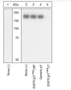
Extracts of A431 cells unstimulated (1) or stimulated with 200 ng/mL EGF for 15 minutes (2-5), were probed with the EGFR [pY1068] antibody following prior incubation with: no peptide (1 and 2), the non-phosphopeptide corresponding to the phosphopeptide immunogen (3), a generic phosphotyrosine-containing peptide (4) or the phosphopeptide immunogen (5). The data show that only the phosphopeptide corresponding to EGFR [pY1068] blocks the antibody signal, demonstrating the specificity of the antibody. The data also show the induction of EGFR [pY1068] phosphorylation by the addition of EGF to this cell system.| Product Name | EGFR (phospho Tyr1068) antibody |
|---|---|
| Antibody Type | Primary Antibodies |
| Product description | Rabbit polyclonal to EGFR ( phospho Y1068 )Epidermal Growth Factor Receptor, (EGFR, a 185 kDa glycoprotein) is a transmembrane tyrosine kinase that regulates a variety of biological responses ranging from mitogenesis to stress signaling. The EGFR consists of a large extracellular domain, a single transmembrane domain and a cytoplasmic domain that exhibits kinase activity. Upon binding of EGF to the extracellular domain, the receptor undergoes dimerization and becomes phosphorylated on several tyrosine residues within the cytoplasmic domain. These events result in EGFR activation and increased tyrosine kinase activity toward a variety of intracellular substrates. Tyrosine 1068 within the cytoplasmic domain of the receptor is an autophosphorylation site that allows binding of Grb2 and activation of the Ras, Raf, ERK1&2 signaling pathway. |
| Immunogen | Synthetic phosphopeptide derived from the region of EGFR that contains tyrosine 1068. |
| Modification | p-Tyr1068 |
| Clonality | Polyclonal |
|---|---|
| Isotype | IgG |
| Host Species | Rabbit |
| Tested Applications | WB |
| WB: 1/1000. Detects a band of approximately 185 kDa. Optimal dilutions/concentrations should be determined by the end user.: |
|
| Species Reactivity | HumanMouseRat |
| Concentration | 1mg/ml |
| Purification | Affinity purified |
| Gene Synonyms | ERBB1 131550 1956 EGFR ERBB mENA P00533 EGFR (phospho Y1068) EGFR (phospho Tyr1068) EGFR (pY1068) EGFR (pTyr1068) EGFR phospho Y1068 EGFR phospho Tyr1068 Phospho EGFR (Y1068) Phospho EGFR (Tyr1068) Phospho EGFR (pY1068) Phospho EGFR (pTyr10 |
|---|---|
| Target | EGFR |
| Tissue Specificity | Specifically recognizes epidermal growth factor receptor phosphorylated at tyrosine 1068. |
| Cellular Localization | Cell Membrane and Secreted |
Application
Extracts of A431 cells unstimulated (1) or stimulated with 200 ng/mL EGF for 15 minutes (2-5), were probed with the EGFR [pY1068] antibody following prior incubation with: no peptide (1 and 2), the non-phosphopeptide corresponding to the phosphopeptide immunogen (3), a generic phosphotyrosine-containing peptide (4) or the phosphopeptide immunogen (5). The data show that only the phosphopeptide corresponding to EGFR [pY1068] blocks the antibody signal, demonstrating the specificity of the antibody. The data also show the induction of EGFR [pY1068] phosphorylation by the addition of EGF to this cell system.| Positive Control | NIH3T3 cells expressing human EGFR, A431 cells +/- EGF |
|---|---|
| Application Notes | WB: 1/1000. Detects a band of approximately 185 kDa. Optimal dilutions/concentrations should be determined by the end user.: |
| Form | Liquid |
|---|---|
| Storage Instructions | Upon receipt - Keep as concentrated solution. Aliquot and store at -20ºC or below. Avoid freeze-thaw cycles. |
| Storage Buffer | Preservative: 0.05% Sodium Azide; Constituents: PBS, 50% glycerol, 1mg/ml BSA. pH 7.3 |


