Application
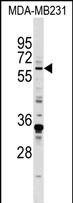
EGR1 Antibody (N-term) western blot analysis in MDA-MB231 cell line lysates (35ug/lane).This demonstrates the EGR1 antibody detected the EGR1 protein (arrow).Application
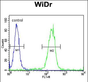
EGR1 Antibody (N-term) flow cytometric analysis of WiDr cells (right histogram) compared to a negative control cell (left histogram).FITC-conjugated goat-anti-rabbit secondary antibodies were used for the analysis.Application
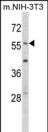
Western blot analysis of EGR1 Antibody (N-term) in mouse NIH-3T3 tissue lysates (35ug/lane). EGR1 (arrow) was detected using the purified Pab.Application
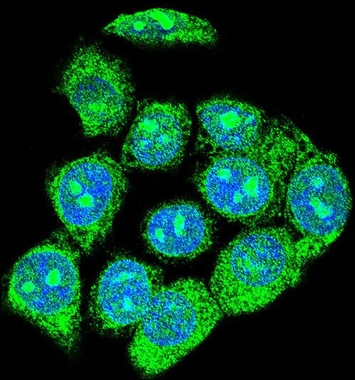
Confocal immunofluorescent analysis of EGR1 Antibody (N-term) with hela cell followed by Alexa Fluor 488-conjugated goat anti-rabbit lgG (green). DAPI was used to stain the cell nuclear (blue).| Product Name | EGR1 antibody, N-term |
|---|---|
| Antibody Type | Primary Antibodies |
| Product description | Rabbit polyclonal to EGR1 (N-term)The protein encoded by this gene belongs to the EGR family of C2H2-type zinc-finger proteins. It is a nuclear protein and functions as a transcriptional regulator. The products of target genes it activates are required for differentitation and mitogenesis. Studies suggest this is a cancer suppresor gene. [provided by RefSeq] |
| Immunogen | KLH conjugated synthetic peptide between 16~45 amino acids from the N-terminal region of human EGR1. |
| Clonality | Polyclonal |
|---|---|
| Isotype | IgG |
| Host Species | Rabbit |
| Tested Applications | FACSICC/IFIHC-PWB |
| ELISA: Use at 1:1,000: |
|
| Species Reactivity | HumanMouse |
| Concentration | 0.2mg/ml |
| Gene Synonyms | Zinc finger protein 225 NGFIA Transcription factor ETR103 EGR1 Nerve growth factor-induced protein A ZNF225 Early growth response protein 1 Nerve growth factorinduced protein A Early growth response protein1 EGR-1 Protein Krox-24 AT225 Protein |
|---|---|
| Target | EGR1 |
| Molecular Weight(MW) | 57.507 kDa (note) |
Application
EGR1 Antibody (N-term) western blot analysis in MDA-MB231 cell line lysates (35ug/lane).This demonstrates the EGR1 antibody detected the EGR1 protein (arrow).
Application
EGR1 Antibody (N-term) flow cytometric analysis of WiDr cells (right histogram) compared to a negative control cell (left histogram).FITC-conjugated goat-anti-rabbit secondary antibodies were used for the analysis.
Application
Western blot analysis of EGR1 Antibody (N-term) in mouse NIH-3T3 tissue lysates (35ug/lane). EGR1 (arrow) was detected using the purified Pab.
Application
Confocal immunofluorescent analysis of EGR1 Antibody (N-term) with hela cell followed by Alexa Fluor 488-conjugated goat anti-rabbit lgG (green). DAPI was used to stain the cell nuclear (blue).| Application Notes | ELISA: Use at 1:1,000: |
|---|
| Form | Liquid |
|---|---|
| Storage Instructions | Store at at 2-8°C for up to 6 months. For long term storage, aliquot and store at -20 °C. Avoid repeated freeze/thaw cycles. |
| Storage Buffer | PBS with 0.09% (W/V) sodium azide |


