Application
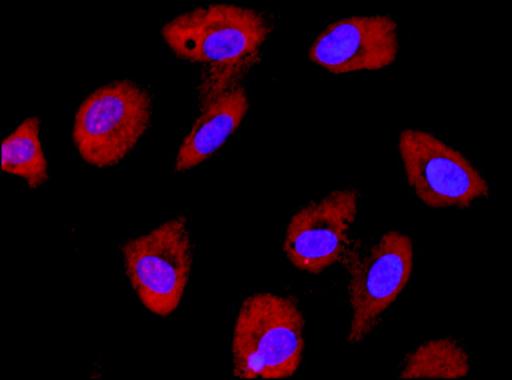
Anti-eIF2Be : Immunofluorescence of HUVEC cells using Anti-eIF2Be (red) at a 1:50 dilution, taken at 40× magnification and nuclear staining with Hoescht 33342 (blue). Yale HTCB IF procedure used.Application
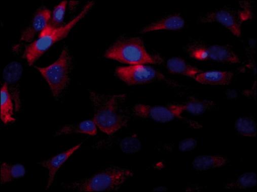
Localization of eIF2Bε in NIH-3T3 cytoplasm. Cells were stained with Rabbit Anti-eIF2Bε at a dilution of 1:1,000 followed by Goat Anti-Rabbit IgG, Cy3 conjugate, and counterstained with DAPI (blue) to stain nuclei (x40 magnification).Application
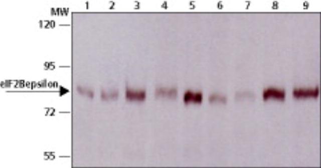
Lysates from various cell lines were separated on SDS-PAGE and probed with 1:500 dilution of Rabbit Anti-eIF2Bε (Cat. No. ). The antibody was developed with Goat Anti-Rabbit IgG, Peroxidase conjugate and a chemiluminiscence substrate. Lanes: 1. 293-T; 2. HeLa; 3. MCF-7; 4. A549; 5. A431; 6. 3T3; 7. C2; 8. RAT1; 9. PC12Application
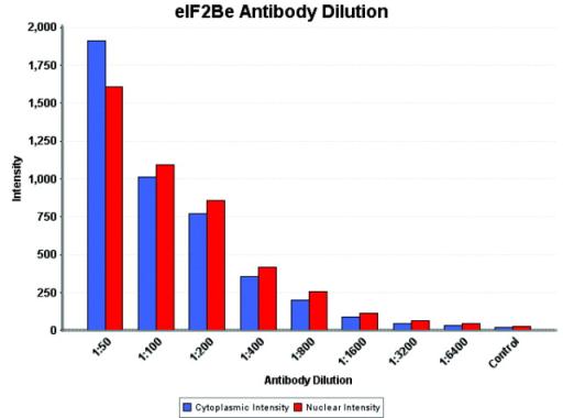
Anti-eIF2Be : Intensity analysis of Anti-eIF2Be staining. Cytoplasmic and nuclear intensity values were obtained at 8 different antibody dilutions (1:50 to 1:6,400) as compared with the negative control.| Product Name | eIF2B antibody |
|---|---|
| Antibody Type | Primary Antibodies |
| Product description | Rabbit polyclonal to eIF2BThis gene encodes one of five subunits of eukaryotic translation initiation factor 2B (EIF2B), a GTP exchange factor for eukaryotic initiation factor 2 and an essential regulator for protein synthesis. Mutations in this gene and the genes encoding other EIF2B subunits have been associated with leukoencephalopathy with vanishing white matter. [provided by RefSeq] |
| Immunogen | Synthetic peptide - Amino acids 50-65 of human eIF2B |
| Clonality | Polyclonal |
|---|---|
| Isotype | IgG |
| Host Species | Rabbit |
| Tested Applications | ICC/IFWB |
| For IF: Use at a dilution of 1:500-1:1,000. For WB: Use at a dilution of 1:500-1:1,000. Optimal dilutions/concentrations should be determined by the end user.: |
|
| Species Reactivity | HumanMouseRat |
| Concentration | 1mg/ml |
| Gene Synonyms | CACH CLE EIF-2B EIF2Bepsilon LVWM EIF 2B EIF2B |
|---|---|
| Target | eIF2B |
Application
Anti-eIF2Be : Immunofluorescence of HUVEC cells using Anti-eIF2Be (red) at a 1:50 dilution, taken at 40× magnification and nuclear staining with Hoescht 33342 (blue). Yale HTCB IF procedure used.
Application
Localization of eIF2Bε in NIH-3T3 cytoplasm. Cells were stained with Rabbit Anti-eIF2Bε at a dilution of 1:1,000 followed by Goat Anti-Rabbit IgG, Cy3 conjugate, and counterstained with DAPI (blue) to stain nuclei (x40 magnification).
Application
Lysates from various cell lines were separated on SDS-PAGE and probed with 1:500 dilution of Rabbit Anti-eIF2Bε (Cat. No. ). The antibody was developed with Goat Anti-Rabbit IgG, Peroxidase conjugate and a chemiluminiscence substrate. Lanes: 1. 293-T; 2. HeLa; 3. MCF-7; 4. A549; 5. A431; 6. 3T3; 7. C2; 8. RAT1; 9. PC12
Application
Anti-eIF2Be : Intensity analysis of Anti-eIF2Be staining. Cytoplasmic and nuclear intensity values were obtained at 8 different antibody dilutions (1:50 to 1:6,400) as compared with the negative control.| Application Notes | For IF: Use at a dilution of 1:500-1:1,000. For WB: Use at a dilution of 1:500-1:1,000. Optimal dilutions/concentrations should be determined by the end user.: |
|---|
| Form | Liquid |
|---|---|
| Storage Instructions | Keep as concentrated solution. Store at 4ºC short term. For extended storage aliquot and store at -20ºC or below. Avoid freeze-thaw cycles. |
| Storage Buffer | PBS, pH7.4 containg 0.1% sodium azide |


