Application
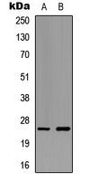
Western blot analysis of Ephrin A5 expression in HeLa (A); NIH3T3 (B) whole cell lysates.Application
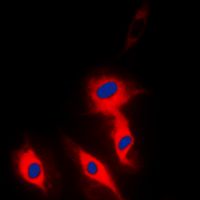
Immunofluorescent analysis of Ephrin A5 staining in Hela cells. Formalin-fixed cells were permeabilized with 0.1% Triton X-100 in TBS for 5-10 minutes and blocked with 3% BSA-PBS for 30 minutes at room temperature. Cells were probed with the primary antibody in 3% BSA-PBS and incubated overnight at 4 deg C in a hidified chamber. Cells were washed with PBST and incubated with a DyLight 594-conjugated secondary antibody (red) in PBS at room temperature in the dark. DAPI was used to stain the cell nuclei (blue).Application
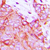
Immunohistochemical analysis of Ephrin A5 staining in human breast cancer formalin fixed paraffin embedded tissue section. The section was pre-treated using heat mediated antigen retrieval with sodium citrate buffer (pH 6.0). The section was then incubated with the antibody at room temperature and detected using an HRP conjugad compact polymer system. DAB was used as the chromogen. The section was then counterstained with haematoxylin and mounted with DPX.WB
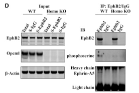
(D) coIP of endogenous EphB2 and endogenous ephrin A5 and immunoblotting of total phosphorylated serine of EphB2 from the hippocampal homogenate of P14 WT and Opcml−/− mice.| Product Name | Ephrin A5 antibody |
|---|---|
| Antibody Type | Primary Antibodies |
| Product description | Rabbit Polyclonal antibody to Ephrin A5Ephrin-A5, a member of the ephrin gene family, prevents axon bundling in cocultures of cortical neurons with astrocytes, a model of late stage nervous system development and differentiation. The EPH and EPH-related receptors comprise the largest subfamily of receptor protein-tyrosine kinases and have been implicated in mediating developmental events, particularly in the nervous system. EPH receptors typically have a single kinase domain and an extracellular region containing a Cys-rich domain and 2 fibronectin type III repeats. The ephrin ligands and receptors have been named by the Eph Nomenclature Committee (1997). Based on their structures and sequence relationships, ephrins are divided into the ephrin-A (EFNA) class, which are anchored to the membrane by a glycosylphosphatidylinositol linkage, and the ephrin-B (EFNB) class, which are transmembrane proteins. The Eph family of receptors are similarly divided into 2 groups based on the similarity of their extracellular domain sequences and their affinities for binding ephrin-A and ephrin-B ligands. [provided by RefSeq, Jul 2008] |
| Immunogen | KLH-conjugated synthetic peptide encompassing a sequence within the center region of human Ephrin A5. The exact sequence is proprietary. |
| Clonality | Polyclonal |
|---|---|
| Isotype | IgG |
| Host Species | Rabbit |
| Tested Applications | FACSICC/IFIHCIHC-PWB |
| FACS:1:200~1:500 ICC/IF:1:200~1:500 IHC:1:50~1:200 IHC-P:1:50~1:200 WB:1:500~1:2000 Notes:Optimal dilutions/concentrations should be determined by the researcher. |
|
| Species Reactivity | BovineChickenHumanMouseRabbitRat |
| Concentration | 1mg/ml |
| Purification | Affinity purified |
| Gene Synonyms | EFL5 LERK7 ephrin-A5 AF1 EPLG7 EFNA5 RAGS GLC1M |
|---|---|
| Target | Ephrin A5 |
| Tissue Specificity | Recognizes endogenous levels of Ephrin A5 protein. |
Application
Western blot analysis of Ephrin A5 expression in HeLa (A); NIH3T3 (B) whole cell lysates.
Application
Immunofluorescent analysis of Ephrin A5 staining in Hela cells. Formalin-fixed cells were permeabilized with 0.1% Triton X-100 in TBS for 5-10 minutes and blocked with 3% BSA-PBS for 30 minutes at room temperature. Cells were probed with the primary antibody in 3% BSA-PBS and incubated overnight at 4 deg C in a hidified chamber. Cells were washed with PBST and incubated with a DyLight 594-conjugated secondary antibody (red) in PBS at room temperature in the dark. DAPI was used to stain the cell nuclei (blue).
Application
Immunohistochemical analysis of Ephrin A5 staining in human breast cancer formalin fixed paraffin embedded tissue section. The section was pre-treated using heat mediated antigen retrieval with sodium citrate buffer (pH 6.0). The section was then incubated with the antibody at room temperature and detected using an HRP conjugad compact polymer system. DAB was used as the chromogen. The section was then counterstained with haematoxylin and mounted with DPX.
WB
(D) coIP of endogenous EphB2 and endogenous ephrin A5 and immunoblotting of total phosphorylated serine of EphB2 from the hippocampal homogenate of P14 WT and Opcml−/− mice.| Application Notes | FACS:1:200~1:500 ICC/IF:1:200~1:500 IHC:1:50~1:200 IHC-P:1:50~1:200 WB:1:500~1:2000 Notes:Optimal dilutions/concentrations should be determined by the researcher. |
|---|
| Form | Liquid |
|---|---|
| Storage Instructions | Shipped at 4 C. Upon delivery aliquot and store at -20 C for one year. Avoid freeze/thaw cycles. |
| Storage Buffer | Liquid in 0.42% Potassium phosphate; 0.87% Sodium chloride; pH 7.3; 30% glycerol; and 0.01% sodium azide. |


