Application
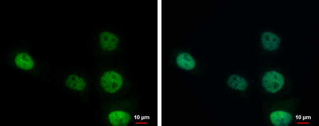
ERCC1 antibody detects ERCC1 protein at nucleus by immunofluorescent analysis. Sample: A431 cells were fixed in 4% paraformaldehyde at RT for 15 min. Green: ERCC1 protein stained by ERCC1 antibody diluted at 1:1000. Blue: Hoechst 33342 staining.Application
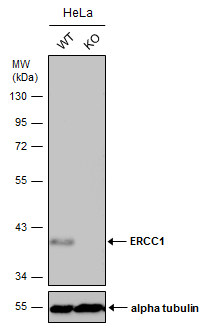
Wild-type (WT) and ERCC1 knockout (KO) HeLa cell extracts (30 µg) were separated by 10% SDS-PAGE, and the membrane was blotted with ERCC1 antibody diluted at 1:1000. The HRP-conjugated anti-rabbit IgG antibody was used to detect the primary antibody.Application
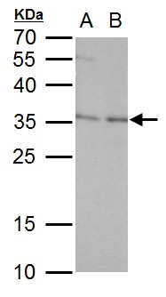
ERCC1 antibody detects ERCC1 protein by western blot analysis. A. 30 μg PC-12 whole cell lysate/extract B. 30 μg Rat2 whole cell lysate/extract 12 % SDS-PAGE ERCC1 antibody dilution: 1:1000Application
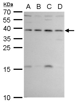
ERCC1 antibody detects ERCC1 protein by western blot analysis. A. 30 μg 293T whole cell lysate/extract B. 30 μg A431 whole cell lysate/extract C. 30 μg HeLa whole cell lysate/extract D. 30 μg HepG2 whole cell lysate/extract 12 % SDS-PAGE ERCC1 antibody dilution: 1:1000Application
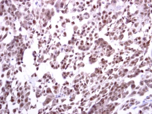
ERCC1 antibody detects ERCC1 protein at cytoplasm and nucleus on human lung carcinoma by immunohistochemical analysis. Sample: Paraffin-embedded human lung carcinoma. ERCC1 antibody diluted at 1:250.| Product Name | ERCC1 antibody |
|---|---|
| Antibody Type | Primary Antibodies |
| Product description | Rabbit Polyclonal antibody to ERCC1 (excision repair cross-complementing rodent repair deficiency, complementation group 1 (includes overlapping antisense sequence))The product of this gene functions in the nucleotide excision repair pathway, and is required for the repair of DNA lesions such as those induced by UV light or formed by electrophilic compounds including cisplatin. The encoded protein forms a heterodimer with the XPF endonuclease (also known as ERCC4), and the heterodimeric endonuclease catalyzes the 5' incision in the process of excising the DNA lesion. The heterodimeric endonuclease is also involved in recombinational DNA repair and in the repair of inter-strand crosslinks. Mutations in this gene result in cerebrooculofacioskeletal syndrome, and polymorphisms that alter expression of this gene may play a role in carcinogenesis. Multiple transcript variants encoding different isoforms have been found for this gene. The last exon of this gene overlaps with the CD3e molecule, epsilon associated protein gene on the opposite strand. [provided by RefSeq] |
| Immunogen | Recombinant protein encompassing a sequence within the center region of human ERCC1. The exact sequence is proprietary. |
| Clonality | Polyclonal |
|---|---|
| Isotype | IgG |
| Host Species | Rabbit |
| Tested Applications | ICC/IFIHC-PWB |
| *Optimal dilutions/concentrations should be determined by the researcher.
: |
|
| Species Reactivity | HumanRat |
| Predicted Reactivity | Mouse, Pig, Chimpanzee, BovineMouse (86%), Pig (87%), Chimpanzee (100%), Bovine (87%) |
| Concentration | 1mg/ml (Please refer to the vial label for the specific concentration) |
| Purification | Affinity purified |
| Gene Synonyms | UV20 RAD10 COFS4 excision repair cross-complementing rodent repair deficiency complementation group 1 (includes overlapping antisense sequence) |
|---|---|
| Target | ERCC1 |
| Molecular Weight(MW) | 33 kDa (note) (2) |
| Cellular Localization | Nucleus |
Application
ERCC1 antibody detects ERCC1 protein at nucleus by immunofluorescent analysis. Sample: A431 cells were fixed in 4% paraformaldehyde at RT for 15 min. Green: ERCC1 protein stained by ERCC1 antibody diluted at 1:1000. Blue: Hoechst 33342 staining.
Application
Wild-type (WT) and ERCC1 knockout (KO) HeLa cell extracts (30 µg) were separated by 10% SDS-PAGE, and the membrane was blotted with ERCC1 antibody diluted at 1:1000. The HRP-conjugated anti-rabbit IgG antibody was used to detect the primary antibody.
Application
ERCC1 antibody detects ERCC1 protein by western blot analysis. A. 30 μg PC-12 whole cell lysate/extract B. 30 μg Rat2 whole cell lysate/extract 12 % SDS-PAGE ERCC1 antibody dilution: 1:1000
Application
ERCC1 antibody detects ERCC1 protein by western blot analysis. A. 30 μg 293T whole cell lysate/extract B. 30 μg A431 whole cell lysate/extract C. 30 μg HeLa whole cell lysate/extract D. 30 μg HepG2 whole cell lysate/extract 12 % SDS-PAGE ERCC1 antibody dilution: 1:1000
Application
ERCC1 antibody detects ERCC1 protein at cytoplasm and nucleus on human lung carcinoma by immunohistochemical analysis. Sample: Paraffin-embedded human lung carcinoma. ERCC1 antibody diluted at 1:250.| Positive Control | 293T , A431 , HeLa , HepG2 , PC-12 , Rat2 , human ERCC1-transfected 293T |
|---|---|
| Application Notes | *Optimal dilutions/concentrations should be determined by the researcher.
: |
| Form | Liquid |
|---|---|
| Storage Instructions | Keep as concentrated solution. Aliquot and store at -20ºC or below. Avoid multiple freeze-thaw cycles. |
| Storage Buffer | 1XPBS, 20% Glycerol (pH7). 0.025% ProClin 300 was added as a preservative. |


