Application
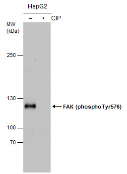
Untreated (–) and treated (+) HepG2 whole cell extracts (30 μg) were separated by 5% SDS-PAGE, and the membrane was blotted with FAK (phospho Tyr576) antibody diluted at 1:1000. (CIP: Calf-intestinal alkaline phosphatase)Application
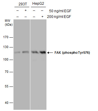
FAK (phospho Tyr576) antibody detects FAK (phospho Tyr576) protein by western blot analysis. Left: Untreated (-) and EGF-treated (+, 50 ng/ml for 30 min) 293T whole cell extracts (30 μg) were separated by 7.5% SDS-PAGE. Right: Untreated (-) and EGF-treated (+, 200 ng/ml for 30 min) HepG2 whole cell extracts (30 μg) were separated by 7.5% SDS-PAGE. The membranes were blotted with FAK (phospho Tyr576) antibody diluted at 1:3000.Application
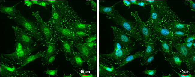
FAK (phospho Tyr576) antibody detects FAK (phospho Tyr576) protein at cell membrane and nucleus by immunofluorescent analysis. Sample: SK-N-SH cells were fixed in 4% paraformaldehyde at RT for 15 min. Green: FAK (phospho Tyr576) protein stained by FAK (phospho Tyr576) antibody diluted at 1:500. Blue: Hoechst 33342 staining. Scale bar = 10 μm.Application
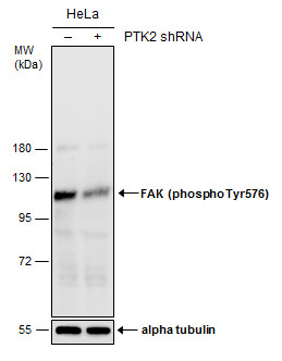
Non-transfected (–) and transfected (+) HeLa whole cell extracts (50 µg) were separated by 7.5% SDS-PAGE, and the membrane was blotted with FAK (phospho Tyr576) antibody diluted at 1:500.| Product Name | FAK (phospho Tyr576) antibody |
|---|---|
| Antibody Type | Primary Antibodies |
| Product description | Rabbit Polyclonal antibody to FAK (phospho Tyr576) (protein tyrosine kinase 2)This gene encodes a cytoplasmic protein tyrosine kinase which is found concentrated in the focal adhesions that form between cells growing in the presence of extracellular matrix constituents. The encoded protein is a member of the FAK subfamily of protein tyrosine kinases but lacks significant sequence similarity to kinases from other subfamilies. Activation of this gene may be an important early step in cell growth and intracellular signal transduction pathways triggered in response to certain neural peptides or to cell interactions with the extracellular matrix. At least four transcript variants encoding four different isoforms have been found for this gene, but the full-length natures of only two of them have been determined. [provided by RefSeq] |
| Immunogen | Carrier-protein conjugated synthetic peptide encompassing a sequence within the center region of Human FAK (phospho Tyr576). The exact sequence is proprietary. |
| Modification | p-Tyr576 |
| Clonality | Polyclonal |
|---|---|
| Isotype | IgG |
| Host Species | Rabbit |
| Tested Applications | ICC/IFWB |
| *Optimal dilutions/concentrations should be determined by the researcher.
: |
|
| Species Reactivity | Human |
| Predicted Reactivity | Mouse, Rat, Xenopus laevis, ChickenMouse (100%), Rat (100%), Xenopus laevis (100%), Chicken (100%) |
| Concentration | 1mg/ml |
| Purification | Affinity purified |
| Gene Synonyms | PPP1R71 FADK protein tyrosine kinase 2 pp125FAK PTK2 FAK FRNK FAK1 p125FAK |
|---|---|
| Target | FAK (phospho Tyr576) |
| Molecular Weight(MW) | 119 kDa (note) (4) |
| Cellular Localization | Cell junction , focal adhesion , Cell membrane; Peripheral membrane protein; Cytoplasmic side |
Application
Untreated (–) and treated (+) HepG2 whole cell extracts (30 μg) were separated by 5% SDS-PAGE, and the membrane was blotted with FAK (phospho Tyr576) antibody diluted at 1:1000. (CIP: Calf-intestinal alkaline phosphatase)
Application
FAK (phospho Tyr576) antibody detects FAK (phospho Tyr576) protein by western blot analysis. Left: Untreated (-) and EGF-treated (+, 50 ng/ml for 30 min) 293T whole cell extracts (30 μg) were separated by 7.5% SDS-PAGE. Right: Untreated (-) and EGF-treated (+, 200 ng/ml for 30 min) HepG2 whole cell extracts (30 μg) were separated by 7.5% SDS-PAGE. The membranes were blotted with FAK (phospho Tyr576) antibody diluted at 1:3000.
Application
FAK (phospho Tyr576) antibody detects FAK (phospho Tyr576) protein at cell membrane and nucleus by immunofluorescent analysis. Sample: SK-N-SH cells were fixed in 4% paraformaldehyde at RT for 15 min. Green: FAK (phospho Tyr576) protein stained by FAK (phospho Tyr576) antibody diluted at 1:500. Blue: Hoechst 33342 staining. Scale bar = 10 μm.
Application
Non-transfected (–) and transfected (+) HeLa whole cell extracts (50 µg) were separated by 7.5% SDS-PAGE, and the membrane was blotted with FAK (phospho Tyr576) antibody diluted at 1:500.| Positive Control | HeLa , 293T , 293T treated 50ng/ml EGF , HepG2 , HepG2 treated 200ng/ml EGF |
|---|---|
| Application Notes | *Optimal dilutions/concentrations should be determined by the researcher.
: |
| Form | Liquid |
|---|---|
| Storage Instructions | Keep as concentrated solution. Aliquot and store at -20ºC or below. Avoid multiple freeze-thaw cycles. |
| Storage Buffer | 1XPBS, 1% BSA, 20% Glycerol (pH7). 0.025% ProClin 300 was added as a preservative. |


