Application
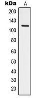
Western blot analysis of Focal Adhesion Kinase (pY925) expression in NIH3T3 pervanadate-treated (A) whole cell lysates.Application
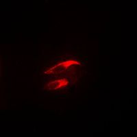
Immunofluorescent analysis of Focal Adhesion Kinase (pY925) staining in NIH3T3 cells. Formalin-fixed cells were permeabilized with 0.1% Triton X-100 in TBS for 5-10 minutes and blocked with 3% BSA-PBS for 30 minutes at room temperature. Cells were probed with the primary antibody in 3% BSA-PBS and incubated overnight at 4 C in a humidified chamber. Cells were washed with PBST and incubated with a DyLight 594-conjugated secondary antibody (red) in PBS at room temperature in the dark. DAPI was used to stain the cell nuclei (blue).| Product Name | FAK (phospho Tyr925) antibody |
|---|---|
| Antibody Type | Primary Antibodies |
| Product description | Rabbit Polyclonal antibody to FAK (phospho Tyr925)This gene encodes a cytoplasmic protein tyrosine kinase which is found concentrated in the focal adhesions that form between cells growing in the presence of extracellular matrix constituents. The encoded protein is a member of the FAK subfamily of protein tyrosine kinases but lacks significant sequence similarity to kinases from other subfamilies. Activation of this gene may be an important early step in cell growth and intracellular signal transduction pathways triggered in response to certain neural peptides or to cell interactions with the extracellular matrix. Several transcript variants encoding different isoforms have been found for this gene, but the full-length natures of only three of them have been determined. [provided by RefSeq, Dec 2010] |
| Immunogen | KLH-conjugated synthetic peptide encompassing a sequence within the C-term region of human Focal Adhesion Kinase. The exact sequence is proprietary. |
| Modification | p-Tyr925 |
| Clonality | Polyclonal |
|---|---|
| Isotype | IgG |
| Host Species | Rabbit |
| Tested Applications | ICC/IFWB |
| WB: 1:500 - 1:1000. ICC/IF: 1:100 - 1:500: |
|
| Species Reactivity | Mouse |
| Concentration | 1mg/ml |
| Purification | Affinity purified |
| Gene Synonyms | FADK PPP1R71 FAK pp125FAK p125FAK PTK2 protein tyrosine kinase 2 FRNK FAK1 |
|---|---|
| Target | FAK (phospho Tyr925) |
| Tissue Specificity | Recognizes endogenous levels of Focal Adhesion Kinase (pY925) protein. |
Application
Western blot analysis of Focal Adhesion Kinase (pY925) expression in NIH3T3 pervanadate-treated (A) whole cell lysates.
Application
Immunofluorescent analysis of Focal Adhesion Kinase (pY925) staining in NIH3T3 cells. Formalin-fixed cells were permeabilized with 0.1% Triton X-100 in TBS for 5-10 minutes and blocked with 3% BSA-PBS for 30 minutes at room temperature. Cells were probed with the primary antibody in 3% BSA-PBS and incubated overnight at 4 C in a humidified chamber. Cells were washed with PBST and incubated with a DyLight 594-conjugated secondary antibody (red) in PBS at room temperature in the dark. DAPI was used to stain the cell nuclei (blue).| Application Notes | WB: 1:500 - 1:1000. ICC/IF: 1:100 - 1:500: |
|---|
| Form | Liquid |
|---|---|
| Storage Instructions | Shipped at 4 °C. Upon delivery aliquot and store at -20 °C for one year. Avoid freeze/thaw cycles. |
| Storage Buffer | Liquid in 0.42% Potassium phosphate; 0.87% Sodium chloride; pH 7.3; 30% glycerol; and 0.01% sodium azide. |


