Application
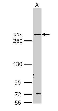
Sample (30 ug of whole cell lysate) A: Molt-4 5% SDS PAGE diluted at 1:1000Application
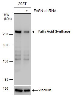
Non-transfected (–) and transfected (+) 293T whole cell extracts (30 µg) were separated by 5% SDS-PAGE, and the membrane was blotted with Fatty Acid Synthase antibody [N1N2], N-term diluted at 1:3000.Application
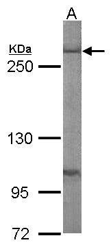
Sample (50 ug of whole cell lysate) A: mouse liver 5% SDS PAGE diluted at 1:1000Application
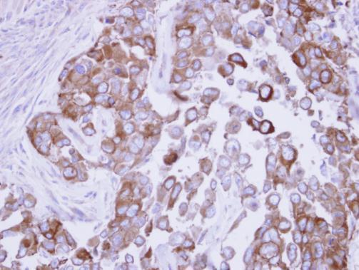
Immunohistochemical analysis of paraffin-embedded OECM1 xenograft, using Fatty Acid Synthase antibody at 1:500 dilution.Application
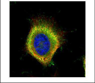
Confocal immunofluorescence analysis (Olympus FV10i) of paraformaldehyde-fixed HeLa, using Fatty Acid Synthase antibody (Green) at 1:500 dilution. Alpha-tubulin filaments were labeled with (Red) at 1:2000.Application
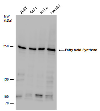
Fatty Acid Synthase antibody detects Fatty Acid Synthase protein by western blot analysis. Various whole cell extracts (30 μg) were separated by 5% SDS-PAGE, and the membrane was blotted with Fatty Acid Synthase antibody diluted by 1:1000.Application
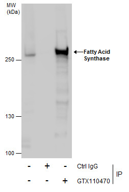
Immunoprecipitation of Fatty Acid Synthase protein from HeLa whole cell extracts using 5 μg of Fatty Acid Synthase antibody [N1N2], N-term . Western blot analysis was performed using Fatty Acid Synthase antibody [N1N2], N-term . EasyBlot anti-Rabbit IgG was used as a secondary reagent.Application
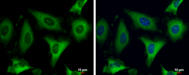
Fatty Acid Synthase antibody [N1N2], N-term detects Fatty Acid Synthase protein at cytoplasm by immunofluorescent analysis. Sample: HeLa cells were fixed in 4% paraformaldehyde at RT for 15 min. Green: Fatty Acid Synthase protein stained by Fatty Acid Synthase antibody [N1N2], N-term diluted at 1:500. Blue: Hoechst 33342 staining. Scale bar = 10 μm.| Product Name | Fatty Acid Synthase antibody , N-term |
|---|---|
| Antibody Type | Primary Antibodies |
| Product description | Rabbit Polyclonal antibody to Fatty Acid Synthase (fatty acid synthase)The enzyme encoded by this gene is a multifunctional protein. Its main function is to catalyze the synthesis of palmitate from acetyl-CoA and malonyl-CoA, in the presence of NADPH, into long-chain saturated fatty acids. In some cancer cell lines, this protein has been found to be fused with estrogen receptor-alpha (ER-alpha), in which the N-terminus of FAS is fused in-frame with the C-terminus of ER-alpha. [provided by RefSeq] |
| Immunogen | Recombinant protein encompassing a sequence within the N-terminus region of human Fatty Acid Synthase. The exact sequence is proprietary. |
| Clonality | Polyclonal |
|---|---|
| Isotype | IgG |
| Host Species | Rabbit |
| Tested Applications | ICC/IFIHC-PIPWB |
| *Optimal dilutions/concentrations should be determined by the researcher.
: |
|
| Species Reactivity | HumanMouse |
| Predicted Reactivity | Rat, BovineRat (85%), Bovine (81%) |
| Concentration | 1mg/ml (Please refer to the vial label for the specific concentration) |
| Purification | Affinity purified |
| Gene Synonyms | FAS antibody MGC14367 antibody MGC15706 antibody OA-519 antibody SDR27X1 antibody FASN antibody "short chain dehydrogenase/reductase family 27X member 1 antibody" fatty acid synthase antibody |
|---|---|
| Molecular Weight(MW) | 273 kDa (note) |
| Cellular Localization | Cytoplasm , Melanosome |
Application
Sample (30 ug of whole cell lysate) A: Molt-4 5% SDS PAGE diluted at 1:1000
Application
Non-transfected (–) and transfected (+) 293T whole cell extracts (30 µg) were separated by 5% SDS-PAGE, and the membrane was blotted with Fatty Acid Synthase antibody [N1N2], N-term diluted at 1:3000.
Application
Sample (50 ug of whole cell lysate) A: mouse liver 5% SDS PAGE diluted at 1:1000
Application
Immunohistochemical analysis of paraffin-embedded OECM1 xenograft, using Fatty Acid Synthase antibody at 1:500 dilution.
Application
Confocal immunofluorescence analysis (Olympus FV10i) of paraformaldehyde-fixed HeLa, using Fatty Acid Synthase antibody (Green) at 1:500 dilution. Alpha-tubulin filaments were labeled with (Red) at 1:2000.
Application
Fatty Acid Synthase antibody detects Fatty Acid Synthase protein by western blot analysis. Various whole cell extracts (30 μg) were separated by 5% SDS-PAGE, and the membrane was blotted with Fatty Acid Synthase antibody diluted by 1:1000.
Application
Immunoprecipitation of Fatty Acid Synthase protein from HeLa whole cell extracts using 5 μg of Fatty Acid Synthase antibody [N1N2], N-term . Western blot analysis was performed using Fatty Acid Synthase antibody [N1N2], N-term . EasyBlot anti-Rabbit IgG was used as a secondary reagent.
Application
Fatty Acid Synthase antibody [N1N2], N-term detects Fatty Acid Synthase protein at cytoplasm by immunofluorescent analysis. Sample: HeLa cells were fixed in 4% paraformaldehyde at RT for 15 min. Green: Fatty Acid Synthase protein stained by Fatty Acid Synthase antibody [N1N2], N-term diluted at 1:500. Blue: Hoechst 33342 staining. Scale bar = 10 μm.| Positive Control | 293T , A431 , HeLa , HepG2 , Molt-4 , Mouse liver |
|---|---|
| Application Notes | *Optimal dilutions/concentrations should be determined by the researcher.
: |
| Form | Liquid |
|---|---|
| Storage Instructions | Keep as concentrated solution. Aliquot and store at -20ºC or below. Avoid multiple freeze-thaw cycles. |
| Storage Buffer | 0.1M Tris, 0.1M Glycine, 10% Glycerol (pH7). 0.01% Thimerosal was added as a preservative. |


