Application
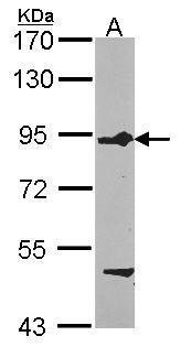
Sample (30 ug of whole cell lysate) A: HCT116 7.5% SDS PAGE diluted at 1:1000Application
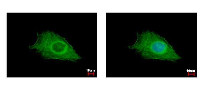
FIG4 antibody detects FIG4 protein at cytoplasm by immunofluorescent analysis. Sample: HeLa cells were fixed in ice-cold MeOH for 5 min. Green: FIG4 protein stained by FIG4 antibody diluted at 1:500. Blue: Hoechst 33342 staining.Application
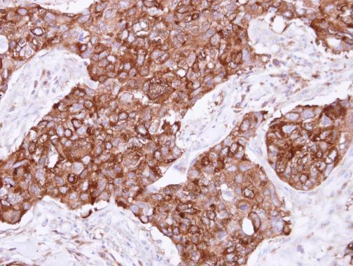
Immunohistochemical analysis of paraffin-embedded human breast cancer, using FIG4 antibody at 1:500 dilution.| Product Name | FIG4 antibody |
|---|---|
| Antibody Type | Primary Antibodies |
| Product description | Rabbit Polyclonal antibody to FIG4 (FIG4 homolog, SAC1 lipid phosphatase domain containing (S. cerevisiae))The protein encoded by this gene belongs to the SAC domain-containing protein gene family. The SAC domain, approximately 400 amino acids in length and consisting of seven conserved motifs, has been shown to possess phosphoinositide phosphatase activity. The yeast homolog, Sac1p, is involved in the regulation of various phosphoinositides, and affects diverse cellular functions such as actin cytoskeleton organization, Golgi function, and maintenance of vacuole morphology. Membrane-bound phosphoinositides function as signaling molecules and play a key role in vesicle trafficking in eukaryotic cells. Mutations in this gene have been associated with Charcot-Marie-Tooth disease, type 4J. [provided by RefSeq] |
| Immunogen | Recombinant protein encompassing a sequence within the center region of human FIG4. The exact sequence is proprietary. |
| Clonality | Polyclonal |
|---|---|
| Isotype | IgG |
| Host Species | Rabbit |
| Tested Applications | ICC/IFIHC-PWB |
| *Optimal dilutions/concentrations should be determined by the researcher.
: |
|
| Species Reactivity | Human |
| Predicted Reactivity | Mouse, Rat, Dog, Pig, Chicken, Rhesus Monkey, Chimpanzee, BovineMouse (95%), Rat (93%), Dog (98%), Pig (97%), Chicken (90%), Rhesus Monkey (99%), Chimpanzee (99%), Bovine (96%) |
| Concentration | 1mg/ml (Please refer to the vial label for the specific concentration) |
| Purification | Affinity purified |
| Gene Synonyms | ALS11 antibody CMT4J antibody KIAA0274 antibody SAC3 antibody dJ249I4.1 antibody FIG4 antibody Sac domain-containing inositol phosphatase 3 antibody SAC domain-containing protein 3 antibody "FIG4 homolog SAC1 domain containing lipid phosphatase an |
|---|---|
| Molecular Weight(MW) | 104 kDa (note) |
| Cellular Localization | Endosome membrane |
Application
Sample (30 ug of whole cell lysate) A: HCT116 7.5% SDS PAGE diluted at 1:1000
Application
FIG4 antibody detects FIG4 protein at cytoplasm by immunofluorescent analysis. Sample: HeLa cells were fixed in ice-cold MeOH for 5 min. Green: FIG4 protein stained by FIG4 antibody diluted at 1:500. Blue: Hoechst 33342 staining.
Application
Immunohistochemical analysis of paraffin-embedded human breast cancer, using FIG4 antibody at 1:500 dilution.| Positive Control | HeLa , HCT116 |
|---|---|
| Application Notes | *Optimal dilutions/concentrations should be determined by the researcher.
: |
| Form | Liquid |
|---|---|
| Storage Instructions | Keep as concentrated solution. Aliquot and store at -20ºC or below. Avoid multiple freeze-thaw cycles. |
| Storage Buffer | 0.1M Tris, 0.1M Glycine, 10% Glycerol (pH7). 0.01% Thimerosal was added as a preservative. |


