Application
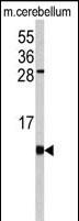
Western blot analysis of FKBP1B antibody (N-term) in mouse cerebellum tissue lysates (35ug/lane). FKBP1B (arrow) was detected using the purified Pab.Application
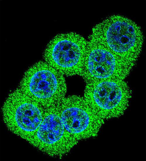
Confocal immunofluorescent analysis of FKBP1B Antibody (N-term) with 293 cell followed by Alexa Fluor 488-conjugated goat anti-rabbit lgG (green). DAPI was used to stain the cell nuclear (blue).Application
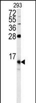
Western blot analysis of FKBP1B antibody (N-term) in 293 cell line lysates (35ug/lane). FKBP1B (arrow) was detected using the purified Pab.Application
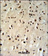
FKBP1B Antibody (N-term) IHC analysis in formalin fixed and paraffin embedded mouse brain tissue followed by peroxidase conjugation of the secondary antibody and DAB staining. This data demonstrates the use of the FKBP1B Antibody (N-term) for immunohistochemistry. Clinical relevance has not been evaluated.WB
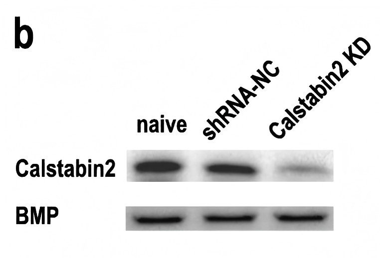
b Western blot showing calstabin2 protein levels in the LA of naive mice, shRNA-NC infected mice, and calstabin2 KD mice. Left: representative Western blot. Right: calstabin2 expression histograms. The y-axis indicates the calstabin2 protein expression level for each condition relative to naive control. The calstabin2 protein level for each condition was normalized to the BMP level. N = 3 for each group. Statistics, One-way ANOVA and Bonferroni post-hoc test. ***p < 0.001 compared with naive and shRNA-NC groups.| Product Name | FKBP1B antibody, N-term |
|---|---|
| Antibody Type | Primary Antibodies |
| Product description | Rabbit polyclonal to FKBP1B (N-term)The protein encoded by this gene is a member of the immunophilin protein family, which play a role in immunoregulation and basic cellular processes involving protein folding and trafficking. This encoded protein is a cis-trans prolyl isomerase that binds the immunosuppressants FK56 and rapamycin. It is highly similar to the FK56-binding protein 1A. Its physiological role is thought to be in excitation-contraction coupling in cardiac muscle. There are two alternatively spliced transcript variants of this gene encoding different isoforms. [provided by RefSeq] |
| Immunogen | The immunogen corresponding to a region within amino acids 16 and 46 of human FKBP1B. |
| Conjugation | Agarose |
| Modification | Acetylated |
| Clonality | Polyclonal |
|---|---|
| Isotype | IgG |
| Host Species | Rabbit |
| Tested Applications | ICC/IFIHC-PWB |
| WB:1:500~1:2000 IHC-P:1:50~1:200 ICC/IF:1:50~1:100 |
|
| Species Reactivity | HumanMouse |
| Concentration | 1mg/ml |
| Gene Synonyms | Peptidylprolyl cistrans isome FKBP-1B Peptidyl-prolyl cis-trans isomerase FKBP1B FKBP1B FKBP1L FKBP12.6 Immunophilin FKBP12.6 12.6 kDa FKBP FKBP9 FK506-binding protein 1B FK506binding protein1B hFKBP12 Peptidylprolyl cistrans isomerase FKBP1B |
|---|---|
| Target | FKBP1B |
| Molecular Weight(MW) | 11.783 kDa (note) |
Application
Western blot analysis of FKBP1B antibody (N-term) in mouse cerebellum tissue lysates (35ug/lane). FKBP1B (arrow) was detected using the purified Pab.
Application
Confocal immunofluorescent analysis of FKBP1B Antibody (N-term) with 293 cell followed by Alexa Fluor 488-conjugated goat anti-rabbit lgG (green). DAPI was used to stain the cell nuclear (blue).
Application
Western blot analysis of FKBP1B antibody (N-term) in 293 cell line lysates (35ug/lane). FKBP1B (arrow) was detected using the purified Pab.
Application
FKBP1B Antibody (N-term) IHC analysis in formalin fixed and paraffin embedded mouse brain tissue followed by peroxidase conjugation of the secondary antibody and DAB staining. This data demonstrates the use of the FKBP1B Antibody (N-term) for immunohistochemistry. Clinical relevance has not been evaluated.
WB
b Western blot showing calstabin2 protein levels in the LA of naive mice, shRNA-NC infected mice, and calstabin2 KD mice. Left: representative Western blot. Right: calstabin2 expression histograms. The y-axis indicates the calstabin2 protein expression level for each condition relative to naive control. The calstabin2 protein level for each condition was normalized to the BMP level. N = 3 for each group. Statistics, One-way ANOVA and Bonferroni post-hoc test. ***p < 0.001 compared with naive and shRNA-NC groups.| Application Notes | WB:1:500~1:2000 IHC-P:1:50~1:200 ICC/IF:1:50~1:100 |
|---|
| Form | Liquid |
|---|---|
| Storage Instructions | Store at at 2-8°C for up to 6 months. For long term storage, aliquot and store at -20 °C. Avoid repeated freeze/thaw cycles. |
| Storage Buffer | PBS with 0.09% (W/V) sodium azide |


