Application
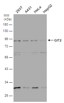
Various whole cell extracts (30 µg) were separated by 10% SDS-PAGE, and the membrane was blotted with GIT2 antibody [N1N3] diluted at 1:1000.Application
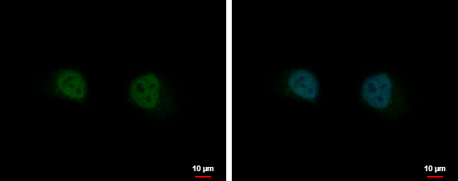
GIT2 antibody [N1N3] detects GIT2 protein at nucleus by immunofluorescent analysis. Sample: HeLa cells were fixed in 4% paraformaldehyde at RT for 15 min. Green: GIT2 protein stained by GIT2 antibody [N1N3] diluted at 1:500. Blue: Hoechst 33342 staining. Scale bar = 10 μm.Application
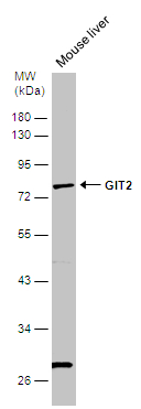
Mouse tissue extract (50 µg) was separated by 10% SDS-PAGE, and the membrane was blotted with GIT2 antibody [N1N3] diluted at 1:1000.| Product Name | GIT2 antibody |
|---|---|
| Antibody Type | Primary Antibodies |
| Product description | Rabbit Polyclonal antibody to GIT2 (G protein-coupled receptor kinase interacting ArfGAP 2)This gene encodes a member of the GIT protein family, which interact with G protein-coupled receptor kinases and possess ADP-ribosylation factor (ARF) GTPase-activating protein (GAP) activity. GIT proteins traffic between cytoplasmic complexes, focal adhesions, and the cell periphery, and interact with Pak interacting exchange factor beta (PIX) to form large oligomeric complexes that transiently recruit other proteins. GIT proteins regulate cytoskeletal dynamics and participate in receptor internalization and membrane trafficking. This gene has been shown to repress lamellipodial extension and focal adhesion turnover, and is thought to regulate cell motility. This gene undergoes extensive alternative splicing to generate multiple isoforms, but the full-length nature of some of these variants has not been determined. The various isoforms have functional differences, with respect to ARF GAP activity and to G protein-coupled receptor kinase 2 binding. [provided by RefSeq] |
| Immunogen | Recombinant protein encompassing a sequence within the center region of human GIT2. The exact sequence is proprietary. |
| Clonality | Polyclonal |
|---|---|
| Isotype | IgG |
| Host Species | Rabbit |
| Tested Applications | ICC/IFWB |
| *Optimal dilutions/concentrations should be determined by the researcher.
: |
|
| Species Reactivity | Human |
| Predicted Reactivity | Mouse, Rat, Xenopus laevis, Chicken, Rhesus Monkey, BovineMouse (96%), Rat (97%), Xenopus laevis (93%), Chicken (96%), Rhesus Monkey (99%), Bovine (99%) |
| Concentration | 1mg/ml |
| Purification | Affinity purified |
| Gene Synonyms | CAT-2 antibody CAT2 antibody DKFZp686G01261 antibody KIAA0148 antibody MGC760 antibody GIT2 antibody "cool-associated tyrosine phosphorylated protein 2 antibody" ARF GTPase-activating protein GIT2 antibody G protein-coupled receptor kinase-interac |
|---|---|
| Molecular Weight(MW) | 85 kDa (note) (8) |
Application
Various whole cell extracts (30 µg) were separated by 10% SDS-PAGE, and the membrane was blotted with GIT2 antibody [N1N3] diluted at 1:1000.
Application
GIT2 antibody [N1N3] detects GIT2 protein at nucleus by immunofluorescent analysis. Sample: HeLa cells were fixed in 4% paraformaldehyde at RT for 15 min. Green: GIT2 protein stained by GIT2 antibody [N1N3] diluted at 1:500. Blue: Hoechst 33342 staining. Scale bar = 10 μm.
Application
Mouse tissue extract (50 µg) was separated by 10% SDS-PAGE, and the membrane was blotted with GIT2 antibody [N1N3] diluted at 1:1000.| Positive Control | A549 , HeLa , HepG2 |
|---|---|
| Application Notes | *Optimal dilutions/concentrations should be determined by the researcher.
: |
| Form | Liquid |
|---|---|
| Storage Instructions | Keep as concentrated solution. Aliquot and store at -20ºC or below. Avoid multiple freeze-thaw cycles. |
| Storage Buffer | 0.1M Tris, 0.1M Glycine, 20% Glycerol (pH7). 0.01% Thimerosal was added as a preservative. |


